
What a year this has been! 2020 has brought so much uncertainty and change into our daily lives. Zoom and Facetime are our new normal, while TikTok keeps a smile on our faces. In what has been a crazy year, we want to look back at how Complete Anatomy has taken you to the next level in your learning.
From listening to our community’s feedback and getting to know you all a little better each year, we’ve managed to pack tons of new learning content into our platform. We’ve empowered you all to succeed even further in your anatomy learning with updates such as a revamped Head & Neck model, 4 new detailed models and an ALL-NEW radiology feature. Let’s dive in a little deeper and see what landed on the platform in 2020.
Radiology – Correlate scans with the 3D model
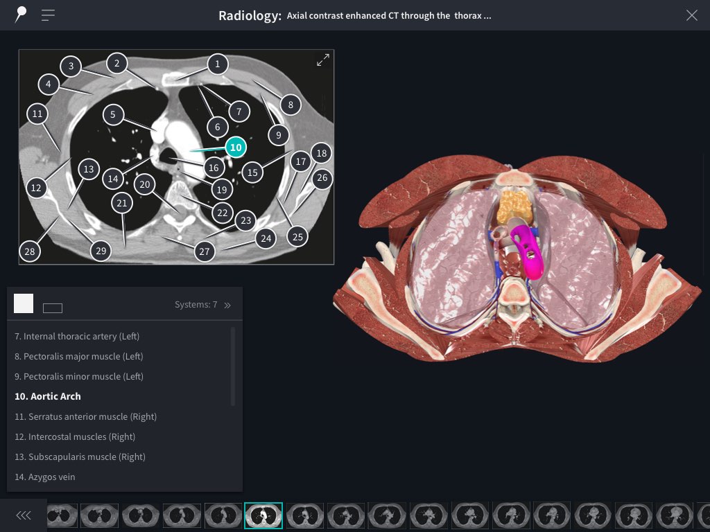
You guys told us time and time again that you want to be able to compare radiology images to the 3D model in order to take your anatomy learning to the next level. Through extensive development and our partnership with Elsevier, we were able to ship our Radiology library this Summer.
Now you can study sequences of images that pass through various anatomical planes, and correlate the pinpointed structures on each image to the 3D model. Containing more detail than any of our competitors in a slick, easy-to-use interface, you are now prepared to enter those later years of anatomy learning.
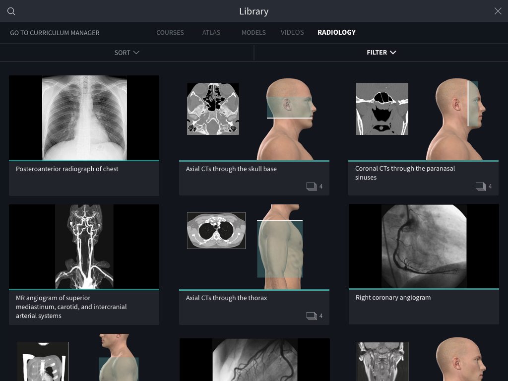
Pssst! Keep an eye out for a brand-new addition to our Radiology library — the upper limb region!
Revamped: Head & Neck model
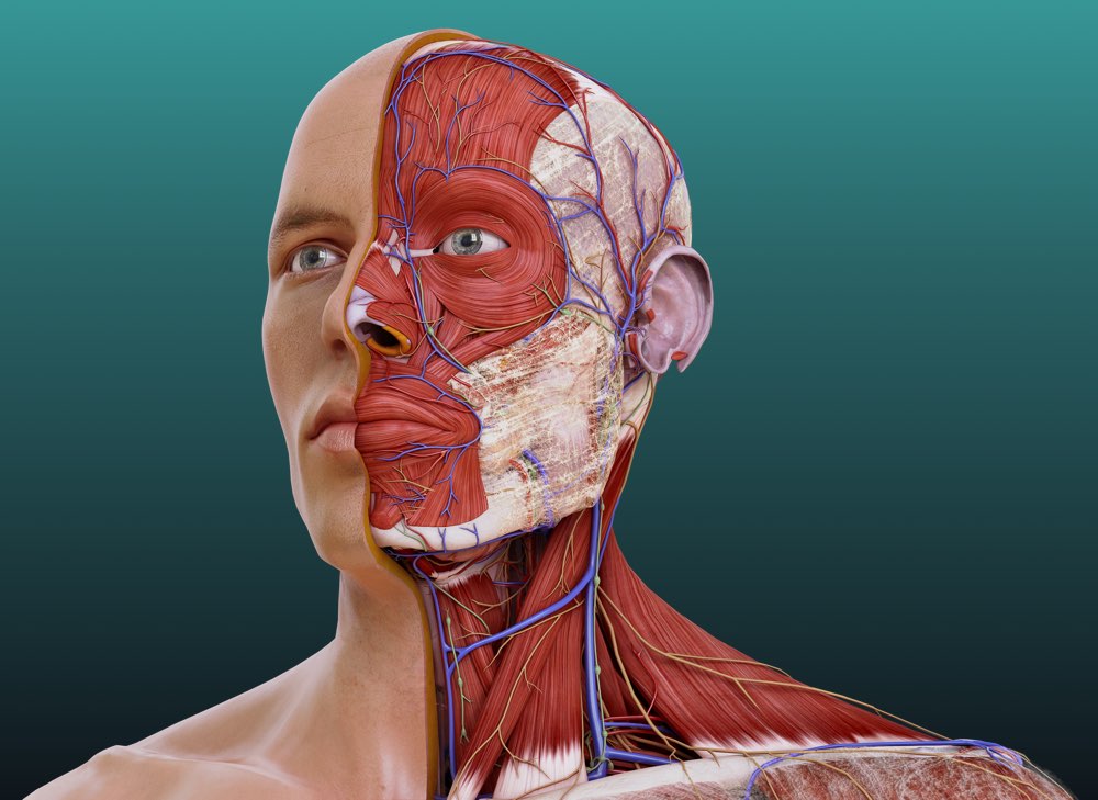
Consistent model updates keep us head and shoulders above the rest, and this one’s no exception! With over 2,000 structures remodeled (700 of which being new), we were delighted to release the result of over 2 years of research and 3D development in 2020.
The skull has been remodeled in its entirety, now with the ability to show all important parts, surfaces, landmarks, and identifiable foramina. We have also introduced, for the first time, craniometrics and sutures to the app.
The muscles of facial expression have been updated, in addition to adding detailed connective tissues such as the superficial musculo-aponeurotic system, a massive WIN for our friends studying facial aesthetics and plastic surgery.
The muscles of the larynx and pharynx also got a revamp, allowing users to fully appreciate the function and structure of breathing and swallowing.
Each of the sense organs have also been remodeled; the eye & the structures of the orbit, the vestibulocochlear organ & bones and ligaments of the inner ear, and finally the tongue, to show each of the muscles which contribute to its function.
Neurovasculature also got a makeover, paying particular attention to the cranial nerves. The pathway of each nerve is now more accurate and easy to follow than ever. The arteries of the internal and external carotids have been modeled in full, in addition to a more accurate representation of the dural venous sinuses and the architecture of the dura mater.
4 NEW detailed models
Cochlea, Photoreceptors, Retinal Layers, and Scalp & Meninges oh my! In 2020 we continued to dive beyond the visible into the innermost workings of the body, with 4 brand-new detailed models.
Cochlea

You wanted more detailed head & neck anatomy and we heard you! ? Explore this sensory organ right down to the cellular level. Combined with our revamped Head & Neck model, you can now follow the transduction of sound from the lymph to the hair cells overlaying the basilar membrane, and along the cochlear nerve to the vestibulocochlear organ and back to the brainstem. Pretty neat, huh?
Retinal Layers
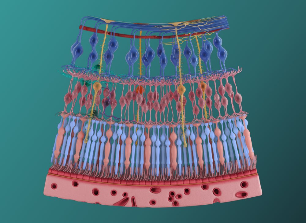
Get an eyeful with our Retinal Layers model. Gain and understanding of the retina and the cells which comprise it, including the retinal ganglion cells, radial glial cells and bipolar neurons. Trace the physiological process of transduction by following the path of light through each of the cell layers, all the way to the photoreceptors.
Photoreceptors
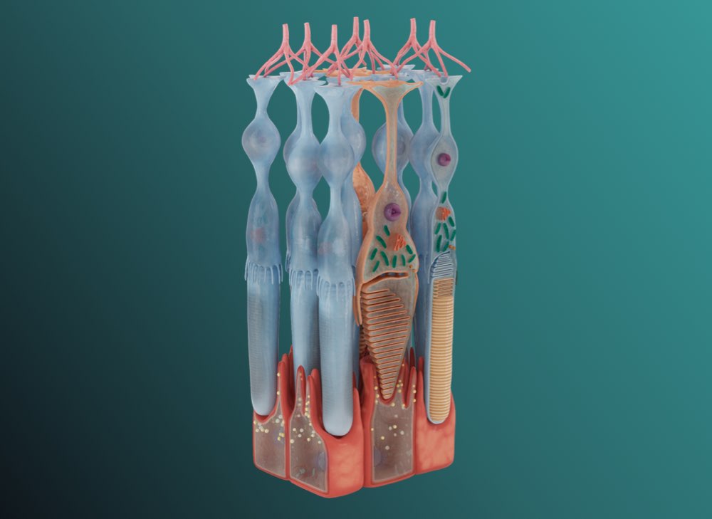
Phototransduction has never been more fun with this incredibly detailed model of the photoreceptor cells (the rods and cones). Explore the unique features of these cells and how they function to absorb light and convert it into an electrical signal.
Scalp & Meninges
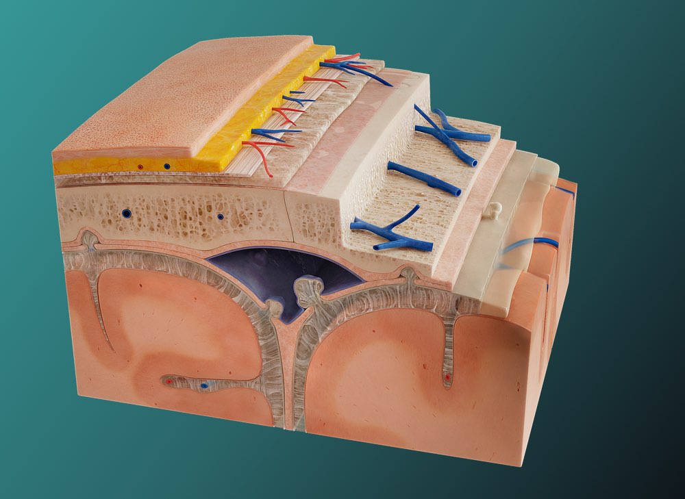
Virtually peel back the five layers of the scalp before moving on to the layers of dura mater. Visually identify the reflections of the meningeal layers and how they enclose the dural venous sinuses and create partitions the brain. The complexities of the interconnected structures in the cranium, including the network of vasculature and folds of each meningeal layer are stunningly depicted in colors and textures that mirror real life anatomy.
And that’s a wrap for 2020!
More than ever, we love and value your continued support throughout 2020. Your feedback has enabled us to consistently deliver top-class learning content that will benefit our community and help anatomy enthusiasts to succeed in classrooms the world over, whether they be real or virtual ones! 2021 will be full of more updates, including 2 new Detailed models in January and an all-new region for our Radiology library. Here’s to 2021 ?. Watch this space!