It’s almost that time again when we launch our biggest update of the year, and this time it’s our most significant product release to date. From a totally revamped Head & Neck model and 4 new detailed models, to an ALL-NEW radiology feature, let’s dive into some of the content landing on the platform this summer.
⭐ NEW: Radiology – Correlate scans with the 3D model
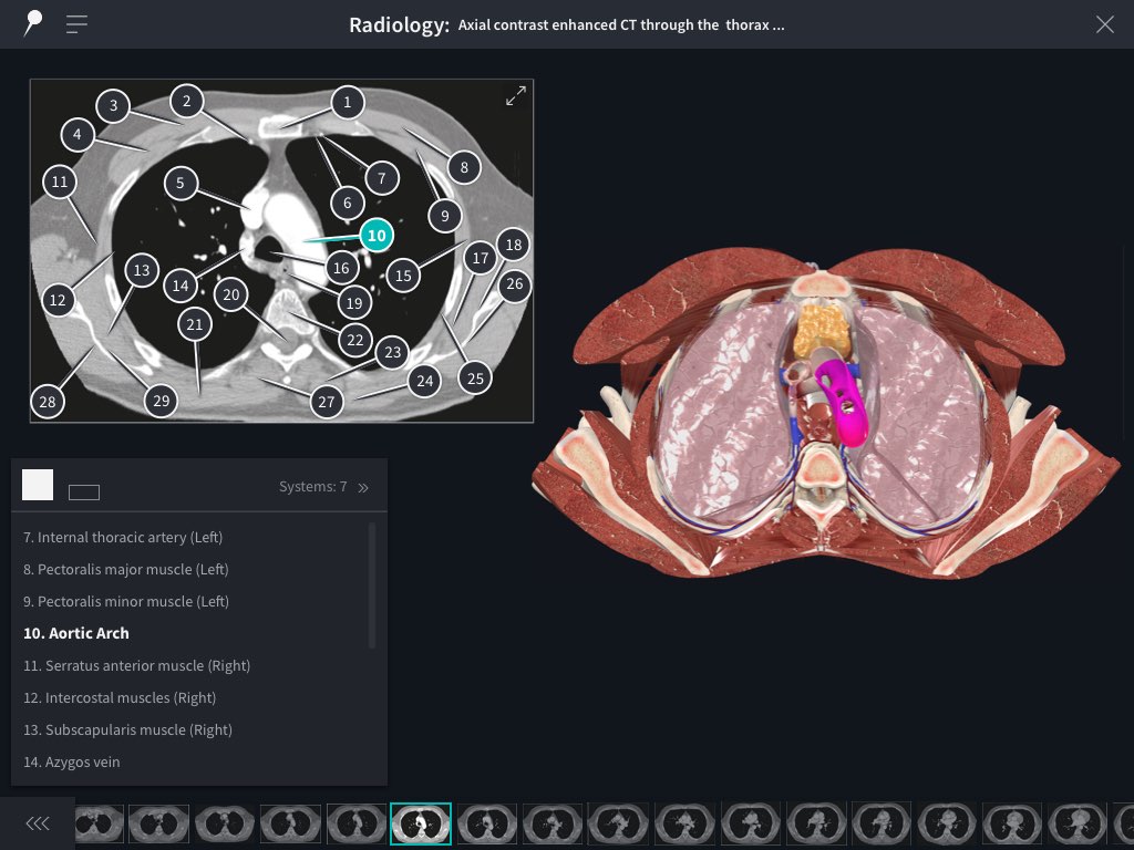
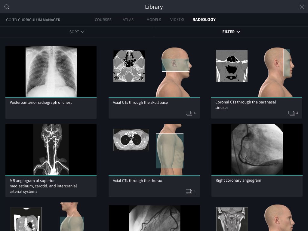
We’ve listened to our customers and we’re excited to present what we’re sure is going to be one of our most popular features. Explore a suite of radiology images from Weir & Abrahams’ whilst gaining a better understanding of them by comparing side-by side with the 3D model. Study sequences of images that pass through various anatomical planes, and correlate the pinpointed structures on each image to the 3D model.
We’ve worked hard to deliver something that’s truly in a league of its own, both in content and design. Here’s just some reasons why our Radiology feature is best in class:
- Depth of information – You’ll find more pinpointed structures on our images than any of our competitors in the 3D space. Furthermore, you can focus on content that matters to you by showing only pins related to certain body systems.
- 3-way-correlation – Identify a structure on-screen by selecting a pin, a structure name, or the 3D model. This flexible approach to learning radiology images something that’s unique to radiology in Complete Anatomy.
- Test your knowledge – Testing yourself before radiology exams has never been easier. Collapse the list to the focused view and then try to name pinned structures, before selecting them to reveal the name. Alternatively, remove pins altogether to simulate a real exam situation.
? REVAMPED: Head & Neck model
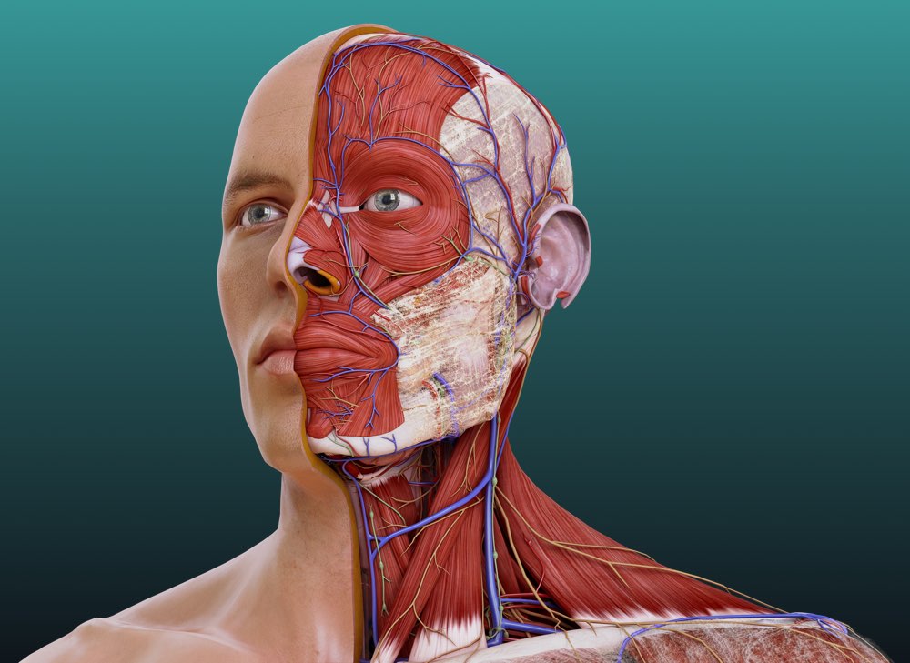
Our latest model update is head and shoulders above the rest – literally! With over 2,000 structures remodeled (700 of which being new), we are delighted to finally release what we’ve been working on for over 2 years.
The skull has been remodeled in its entirety, now with the ability to show all important parts, surfaces, landmarks, and identifiable foramina. We have also introduced, for the first time, craniometrics and sutures to the app.
The muscles of facial expression have been updated, in addition to adding detailed connective tissues such as the superficial musculo-aponeurotic system, a massive win for those studying facial aesthetics and plastic surgery.
The muscles of the larynx and pharynx also got a revamp, allowing users to fully appreciate the function and structure of breathing and swallowing.
Each of the sense organs have also been remodelled; the eye & the structures of the orbit, the vestibulocochlear organ & bones and ligaments of the middle ear, and finally the tongue, to show each of the muscles which contribute to its function.
Neurovasculature also got a makeover, paying particular attention to the cranial nerves. The pathway of each nerve is now more accurate and easy to follow than ever. The arteries of the internal and external carotids have been modeled in full, in addition to a more accurate representation of the dural venous sinuses and the architecture of the dura mater.
4 NEW detailed models
We continue to dive beyond the visible into the innermost workings of the body with 4 brand-new detailed models: Cochlea, Retinal Layers, Photoreceptors, and Scalp & Meninges.
Cochlea

Explore the sensory organ of hearing right down to the cellular level. Combined with our revamped Head & Neck model, you can now follow the transduction of sound from the perilymph to the hair cells overlying the basilar membrane, and along the cochlear nerve to the vestibulocochlear organ and onto the brainstem.
Retinal Layers
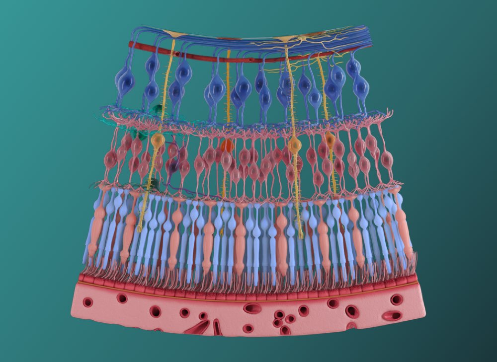
Gain a more in-depth understanding of the retina and the cells which comprise it, including the retinal ganglion cells, radial glial cells and bipolar neurons. Trace the physiological process of transduction by following the path of light through each of the cell layers, all the way to the photoreceptors.
Photoreceptors
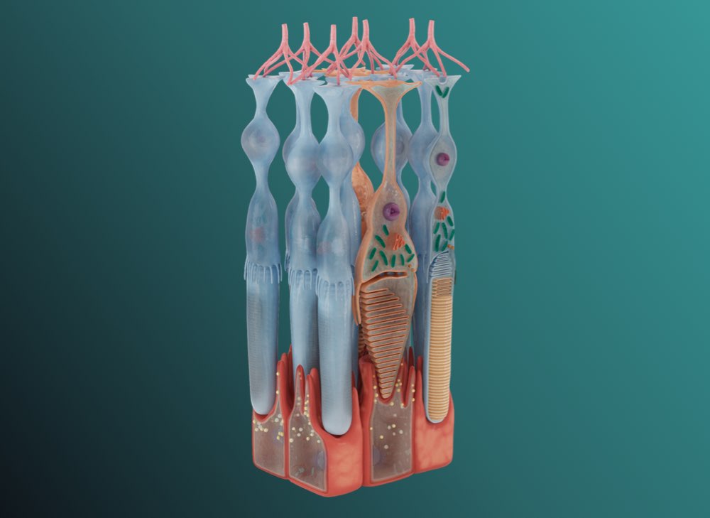
Fully understand the process of phototransduction with this incredibly detailed model of the photoreceptor cells (the rods and cones). Explore the unique features of these cells and how they function to absorb light and convert it into an electrical signal.
Scalp & Meninges
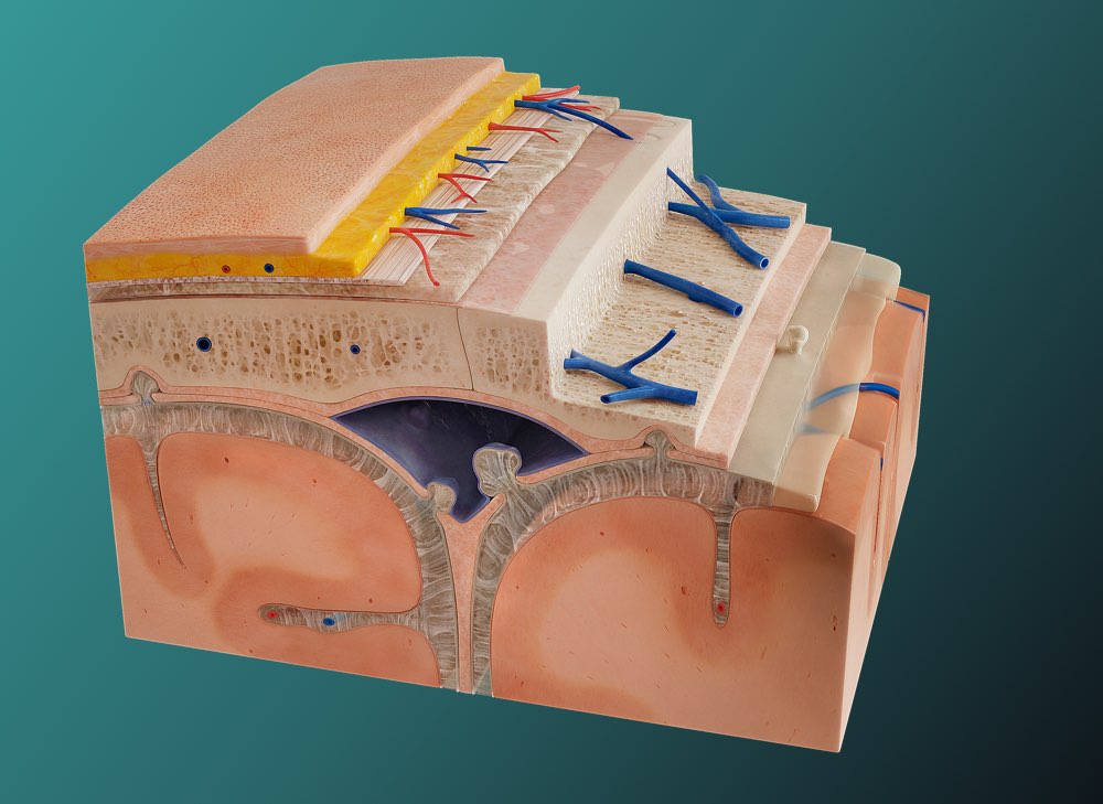
Virtually peel back the five layers of the scalp before moving on to the layers of dura mater. This detailed model illustrates reflections of the meningeal layers and how they enclose the dural venous sinuses and create partitions the brain. The complexities of the interconnected structures in the cranium, including the network of vasculature and folds of each meningeal layer are stunningly depicted in colours and textures that mirror real life anatomy.
We’re working round the clock to deliver our biggest update of the year, and we’re very nearly there. Be sure to opt-in for New Technology Previews in your email preference, and be the first to find out when our summer release lands on the app stores.