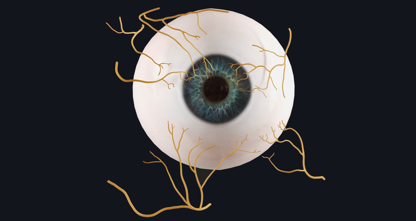
The eye is an amazing and complex structure ?. In fact, the only other organ that’s more complex than is the brain. Incredibly, the eye is made up of over 2 million working parts — the optic nerve alone contains between 770,000 and 1.7 million nerve fibers ?. Read on to learn more about the innervation of the eye…
6 cranial nerves innervate motor, sensory, and autonomic structures of the eye.
Optic Nerve (CN II):
The function of this nerve is purely sensory. It senses incoming light and images displayed on the retina. It then sends this information to the cerebral cortex. It also contributes to changing pupil sizes (autonomic).
Oculomotor Nerve (CN III):
This nerve is responsible for the motor innervation of the majority of extraocular muscles. It also contributes to the constriction of pupils (autonomic). *branches not seen above*
Trochlear Nerve (CN IV):
The trochlear nerve also contributes to the motor innervation of the eye. Of the extraocular muscles, it only innervates the superior oblique muscle.
Trigeminal Nerve (CN V):
Of the 3 branches of the trigeminal nerve, the ophthalmic nerve is involved in sensory innervation of the eye. It works as an afferent part of the corneal and lacrimation reflex.
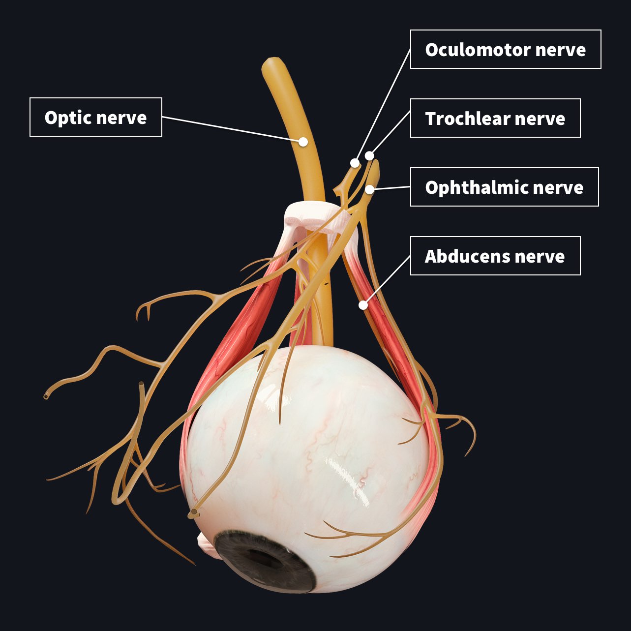
Abducens Nerve (CN VI):
This nerve only innervates one muscle of the eye, the lateral rectus muscle.
Facial Nerve (CN VII):
In the eyes, this nerve is responsible for eye closure and blinking by having motor innervation of the orbicularis oculi muscle. It also has sensory efferent output for the corneal and lacrimation reflex of the eye. *not seen above*
The autonomic nervous system it is divided into…
Sympathetic Nervous System
Pupillary dilation occurs through this system. Its neuron ends in the orbit where it becomes the long ciliary nerve. This nerve innervates the muscles that control pupillary dilation.
Parasympathetic Nervous System
This system causes pupillary constriction. The nerve fibers end at the short ciliary nerves which innervate the sphincter pupillae muscles and cause contraction of the pupil.
Explore the branches and origin paths of the nerves that innervate of all the major organs with our fully interactive gross anatomical model. Try it for FREE today.
If you found this blog post useful, you might also enjoy learning about our most advanced eye model yet.