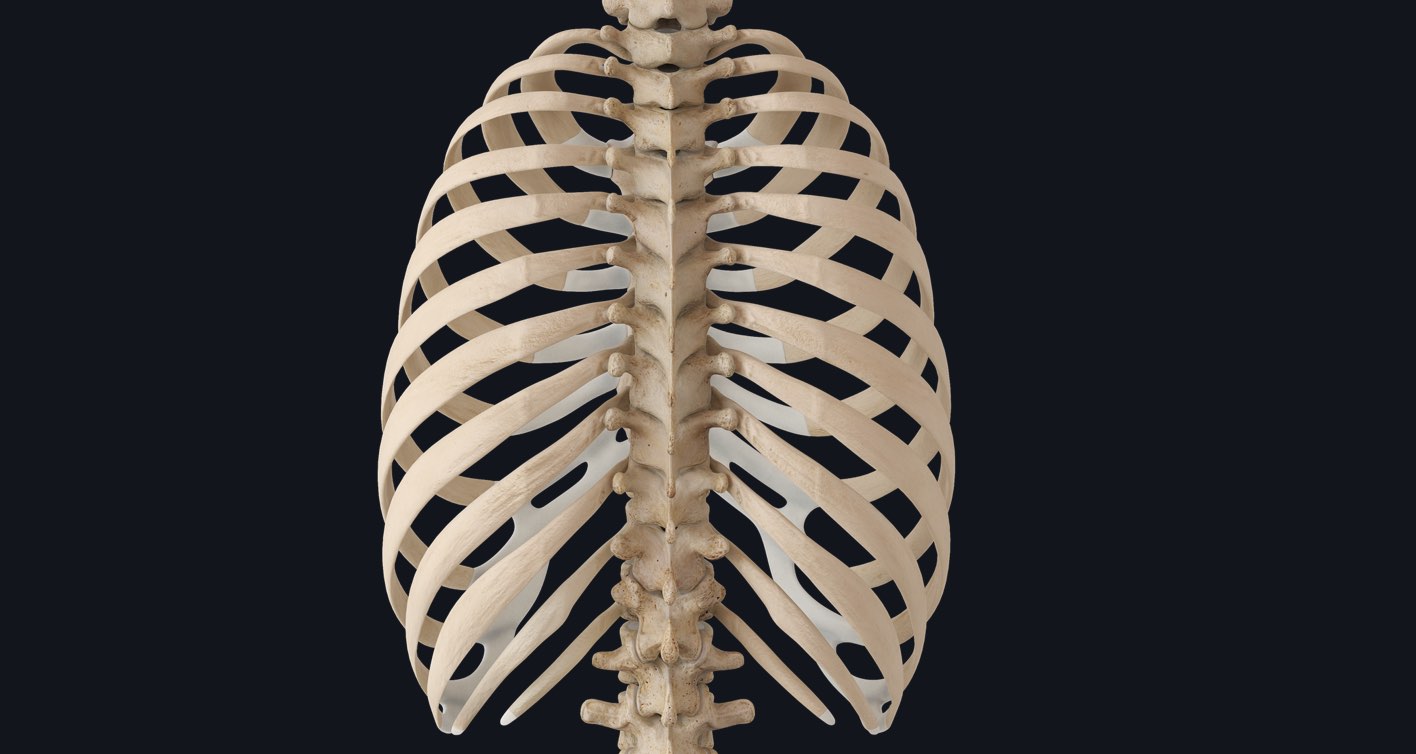
Did you know that without a rib cage, the heart and lungs would not stay in place and it would be virtually impossible to breathe? To say this presents a severe hindrance on the sustainability of life is an understatement! So, let’s look at this vital structure of the thorax in more detail.
The thoracic cage (rib cage) is the osteocartilaginous structure found in the axial skeleton’s thoracic region. It provides the framework for the thoracic wall and protection to organs of the thoracic and upper abdominal regions.
It‘s like a bird cage in appearance and consists of twelve pairs of ribs and the sternum, with the first to tenth ribs connected to the sternum via costal cartilages. The thoracic cage is most extensive, transversely, at the level of the eighth or ninth ribs.
The thoracic cage has openings located superiorly and inferiorly; this allows for communication between the thorax and the body cavities that lie above and below it.
The superior opening is known as the superior thoracic aperture or the thoracic inlet. It is kidney-shaped, smaller than the inferior thoracic aperture, and its outline is defined:
Posteromedially– by the anterior aspect of the T1 vertebra
Laterally– by the internal borders of both the right and left first ribs and their corresponding costal cartilages
Anteromedially– by the superior border of the manubrium of the sternum.
The inferior opening is the inferior thoracic aperture (anatomical thoracic outlet). It is larger than the superior thoracic aperture, and its outline is defined:
Posteromedially– by the anterior aspect of the T12 vertebra
Posterolaterally– by the inferior borders of both the right and left twelfth ribs
Laterally– by the costal cartilages of both the right and left tenth ribs
Anterolaterally- by both the right and left costal margins
Anteromedially– by the xiphisternal joint
These structures provide attachment sites for the diaphragm and a frame to allow adequate space for the lungs to expand. Damage to the rib cage by events such as blunt chest trauma leads to rib fracture. In this situation, the lungs do not expand adequately and can present a paradoxical chest movement known as Flail Chest.
Discover the bones of the thorax, complete with mapping of their parts, surfaces, landmarks and O/I sites. To see how you can get the edge over your class, try Complete Anatomy for FREE today.
