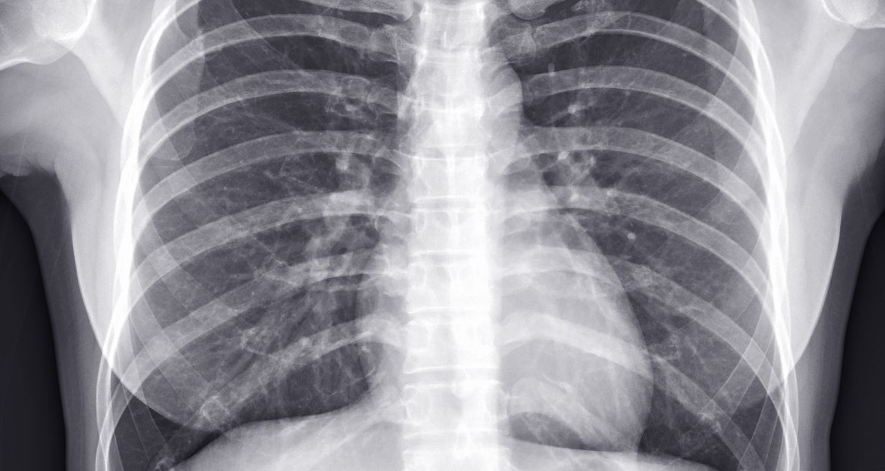
Most of us have seen a chest radiograph before, some may even have had one done. But have you ever wondered what exactly a clinician is looking for when they analyze this black and white image of your insides?
A chest radiograph is usually taken to investigate a suspected pathology in the thorax or upper abdomen. The image produced allows visualization of the bones ?of the thoracic cage, back and part of the upper limbs, as well as some soft tissues such as the lungs ?️and heart ❤️
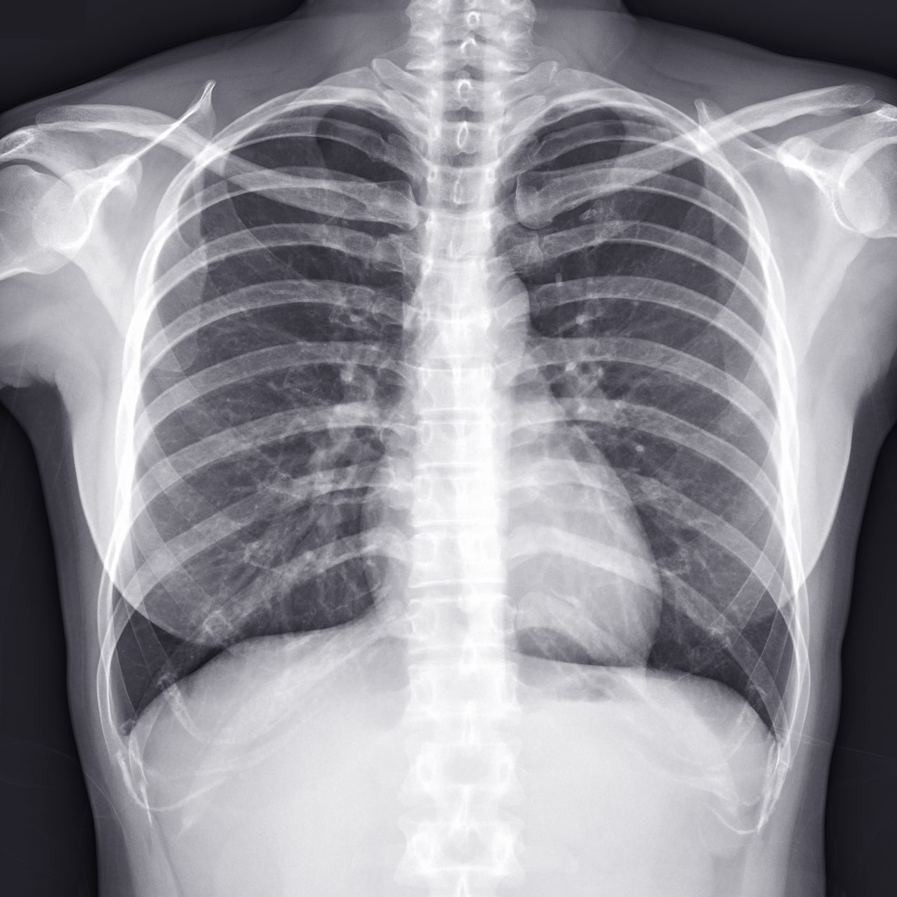
Here is a handy guide that clinicians use to help them through the assessment of a chest radiograph. See if you can make out the structures in the above radiograph using the labelled 3D model in the next image to help you.
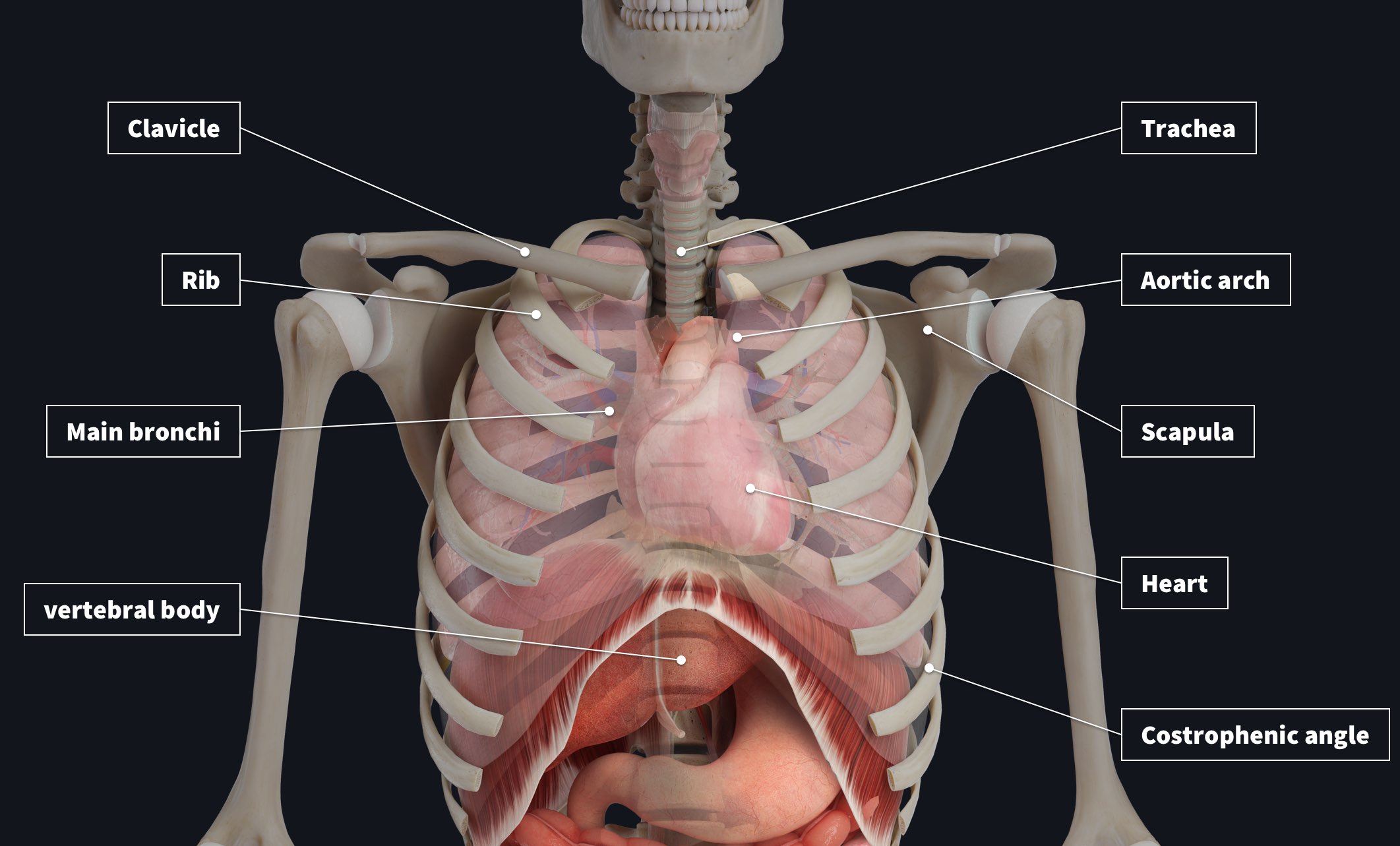
- A: Airway – the trachea should be visible and central as it travels down the neck and the main bronchi should be visible either side of the mediastinum.
- B: Breathing – both lungs should fill the chest cavity; they should have sharp costophrenic angles (the lateral angle). An area of opacity may indicate a pathology.
- C: Circulation – the heart should make up approximately 50% of the diameter of the thorax and most of it should be visible to the left of the midline, the aortic arch should also protrude to the left, and the bronchial vessels should be visible on close inspection.
- D: Disability – follow the course of each rib to see if there are any fractures. The clavicles, scapulae and vertebral bodies should also be reviewed.
- E: Everything Else! – this is a reminder to take a step back and look for things that you might not expect, such as foreign objects that might have been inhaled or swallowed, calcifications or masses, etc.
A radiograph is produced by shooting x-rays through the tissues and recording how many pass through to the other side of the patient. Some tissues will absorb more of these x-rays than others. A film exposed to a large number of x-ray beams will appear black, bones absorb quite a few x rays and that is why they appear white on the film.
Easily import clinical imagery such as MRIs and radiographs and correlate what you see with the 3D model. Try it for FREE today.