Abrahams’ Clinical Human Anatomy

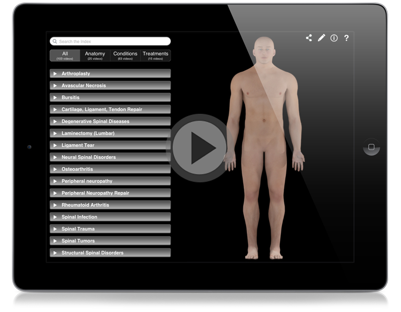
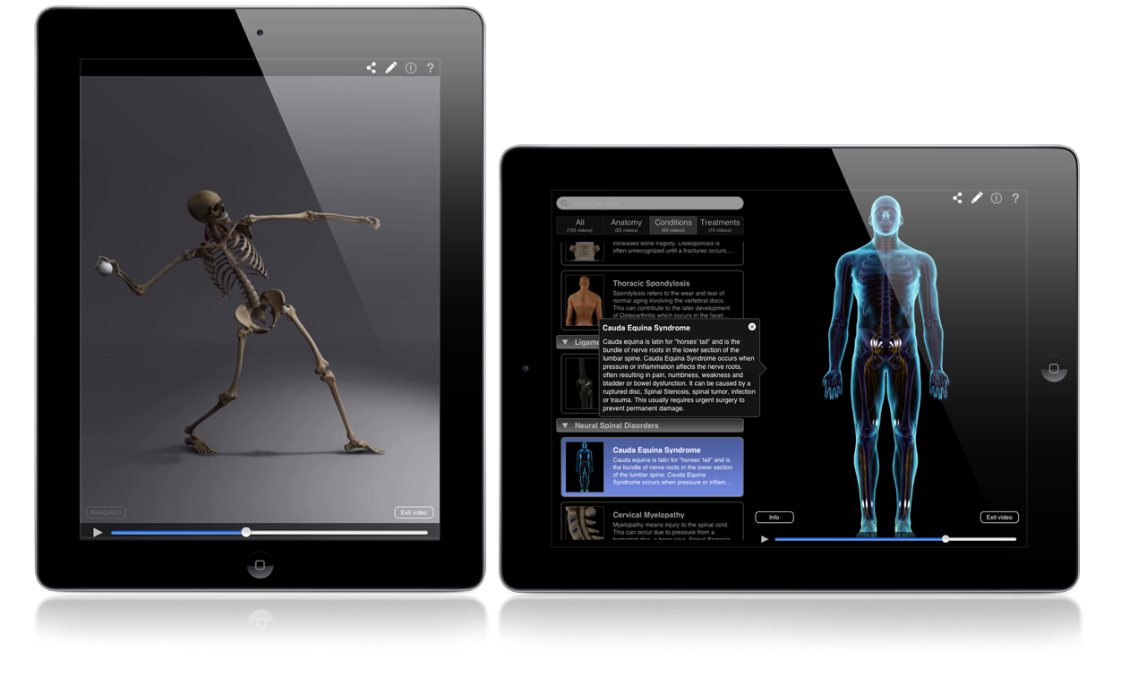
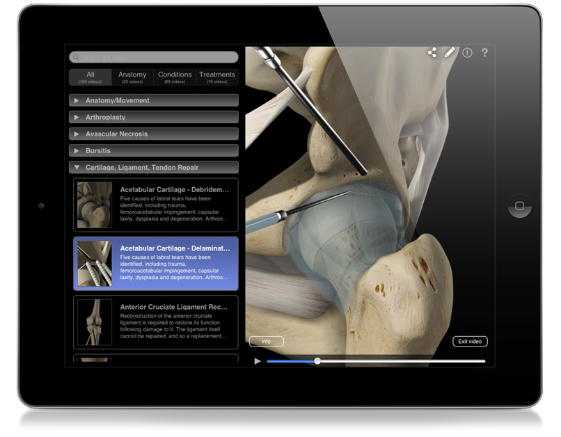
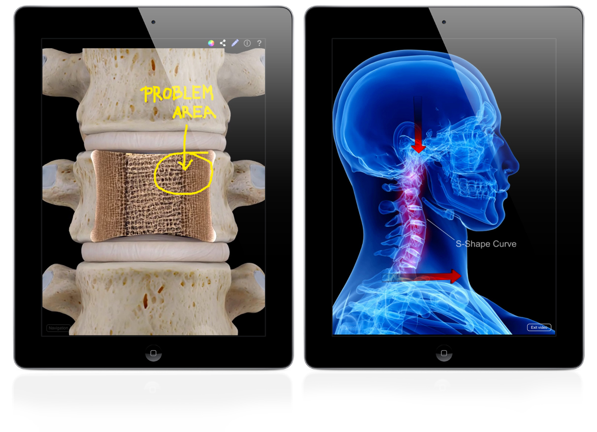
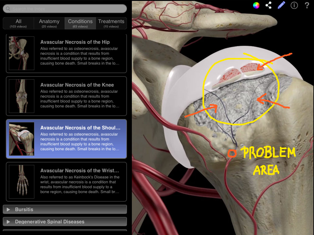
Abrahams’ Clinical Human Anatomy course includes 500 clinical images collected by the leading international anatomist Professor Peter Abrahams throughout his career. This interactive lecture series includes dynamic Screens covering essential clinical conditions which help you gain a better understanding of human anatomy by applying it to clinical practice. Correlate anatomy to clinical practice with a wealth of medical images, including X-RAY, MRI, CT, endoscopic, laparoscopic, cadaveric, ultrasound and clinical images, and explore pathologies in all regions of the body. The detailed model helps you to view the body in a more dynamic way to aid your understanding of anatomical relationships, and the excellent medical images allow you to master essential clinical conditions that every physician should know. The images utilized in the course screens correlate to the published Elsevier text McMinn & Abrahams’ Clinical Atlas of Human Anatomy.

Kinesiology Taping

This course looks at the benefits of kinesiology taping for the lower limb and pelvis. In this course, Aubrey details the different taping strategies for individual anatomical structures, and shows the different therapeutic techniques for specific injuries and dysfunctions. The course is supplemented with videos, detailed descriptions and rationale for the application of each taping procedure.

Point of Care Ultrasound

A supplemental anatomy course introducing the diagnostic imaging modality of ultrasound. Throughout this course the fundamental use and clinical application of ultrasound as a diagnostic tool will be explored through seven key examinations. In this course an overview of an e-FAST, AAA, Renal, Biliary, DVT, Basic Echo, and Lung scan will all be presented. Each scanning technique will introduce a relevant clinical case and guide course participants through the various relevant locations of interest and clinical concern.

Cadaveric Lab: Skeletal and Muscular Systems

A companion course to traditional anatomy lectures featuring detailed photographs of professionally dissected cadaveric tissue. The photographic library represents a comprehensive look at the human body through all body regions and systems. The course is designed to better prepare students in how to identify anatomical structures in embalmed human cadaveric specimens in either dissection or prosection based courses.

Undergraduate Human Anatomy

Undergraduate Human Anatomy is an interactive lecture based course studying terminology, tissues, skeleton, muscles, blood vessels, and nerves of the seven regions of the body. The regions of interest include: Back, Lower Limb, Upper Limb, Head/Neck, Thorax, Abdomen, and Pelvis. The focus of this course we will be to introduce many of the underlying concepts and principles related to human gross anatomy as appropriate for an undergraduate level of education.

Orthopedic Patient Education

About

About

Orthopedic Patient Education is an animation based patient education app for healthcare professionals to effectively communicate anatomy, conditions and treatments to patients. The app covers a variety of conditions such as joint diseases and disorders, as well as joint physiology. *The Consultation feature is currently only available in the iPad version, but will be added to the Mac version in the near future.
3D Technology and Stunning Graphics

Browse

Included within the app are over 100 beautifully detailed animations that allow users to effortlessly search via category. Further description of each animation is also given in a quick and easy to use reference index.
3D Technology and Stunning Graphics

Content

This app draws upon animations from 3D4Medical’s Body Region Pro series of apps, as well as additional animations unavailable anywhere else. All of these animations have been brought together to make for a comprehensive repository for orthopedic conditions and treatments. Additional animations will be added to the app on a continual basis.
3D Technology and Stunning Graphics

Consult

The app allows healthcare professionals to pause and then draw on any animation with the pen tool to highlight and annotate specific images within an animation. These images created during their consultation can also be sent to the patient via email. *The Consultation feature is currently only available in the iPad version, but will be added to the Mac version in the near future.