Complete Anatomy
Abrahams’ Clinical Human Anatomy
About this course
By Dr. Peter Abrahams
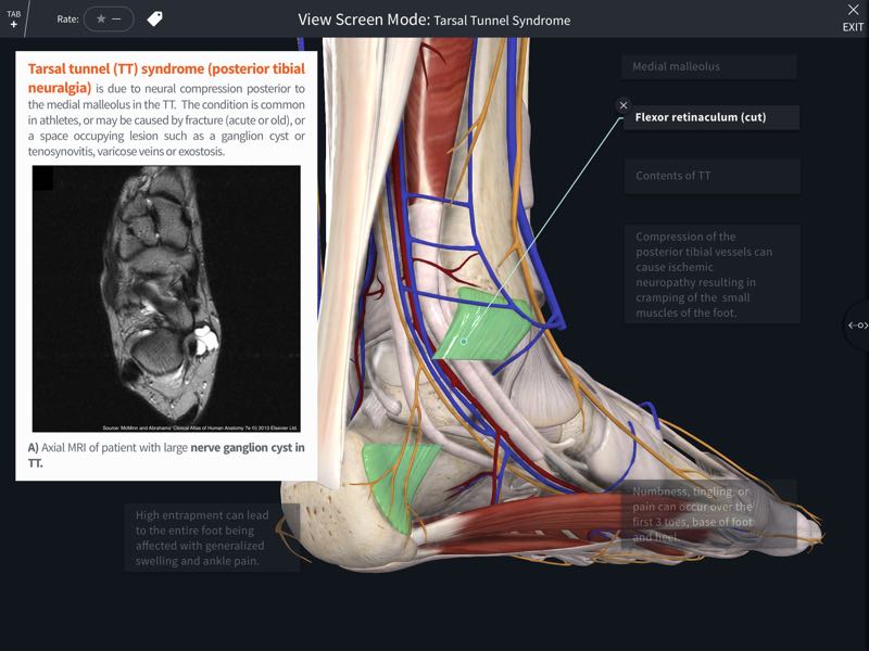
Abrahams’ Clinical Human Anatomy course includes 500 clinical images collected by the leading international anatomist Professor Peter Abrahams throughout his career. This interactive lecture series includes dynamic Screens covering essential clinical conditions which help you gain a better understanding of human anatomy by applying it to clinical practice. Correlate anatomy to clinical practice with a wealth of medical images, including X-RAY, MRI, CT, endoscopic, laparoscopic, cadaveric, ultrasound and clinical images, and explore pathologies in all regions of the body. The detailed model helps you to view the body in a more dynamic way to aid your understanding of anatomical relationships, and the excellent medical images allow you to master essential clinical conditions that every physician should know. The images utilized in the course screens correlate to the published Elsevier text McMinn & Abrahams’ Clinical Atlas of Human Anatomy.
Learning Outcomes
On completion of this course you will be able to correlate human anatomy to essential clinical conditions.
Author
Peter Abrahams, MBBS FRCS(ED) FRCR DO(Hon) FHEA, has been a Professor at St. George's University, Grenada, since 1993. Having written 'Clinical Anatomy of Practical Procedures' with Webb in 1973, which led to him being awarded a British Fulbright Scholarship to the University of Iowa in 1975, he then held the position of Clinical Anatomist at UCL (London), followed by the same post at The University of Cambridge, before becoming the Professor of Clinical Anatomy at the Kigezi International School of Medicine. Having worked as an educational and anatomical consultant for the WHO in Geneva, he also set up a school in Beersheba and has lectured doctors and surgeons around the world. He has published 18 student textbooks on clinical anatomy and radiology, which have been published in over 70 languages. His major works include the McMinn 'Atlas of Human Anatomy', now in its 6th edition; and 'Imaging Atlas of Human Anatomy' with Weir, currently in its 4th edition. In 2006, the AACA recognized Dr. Abrahams as an international clinical anatomist, teacher, author and family doctor, and awarded him with 'Honored Member Status' in the AACA. In 2011, Dr. Abrahams was awarded the National Teaching Fellowship from the HEA, the highest teaching & educational award in UK universities.