Complete Anatomy
Point of Care Ultrasound

About this course
By Dr. Alan Detton, Khanh Nguyen & David Resuehr
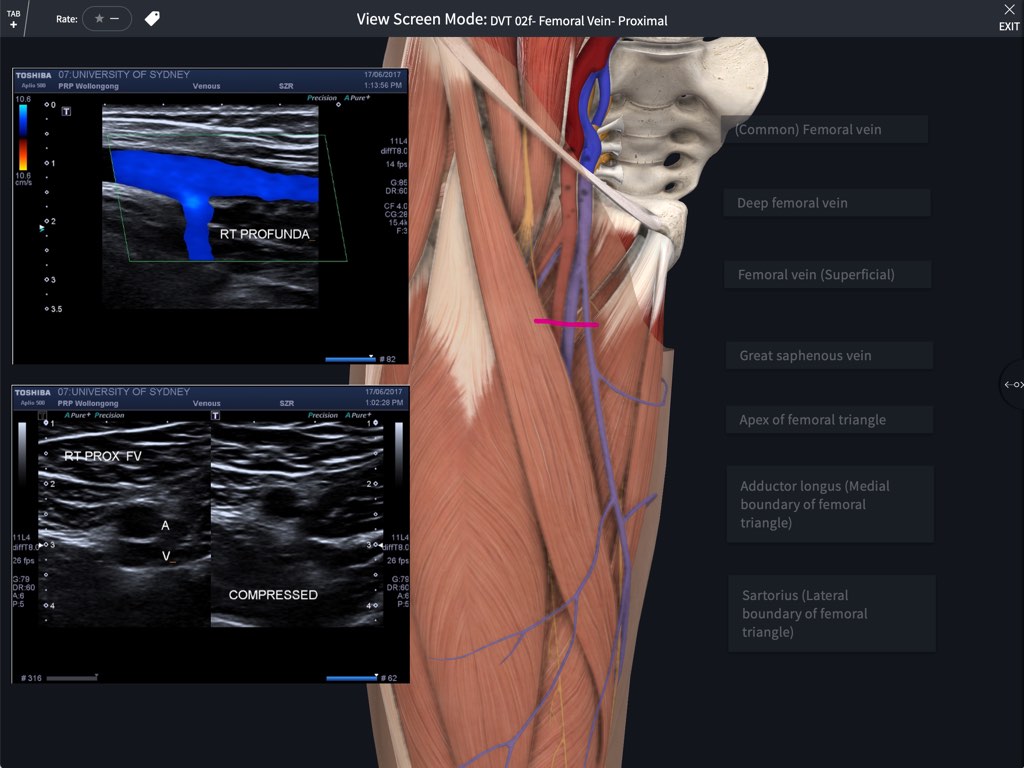
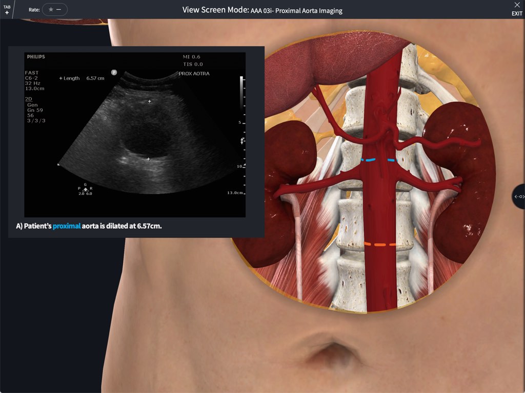
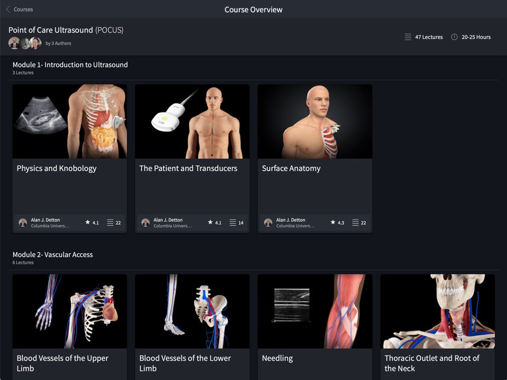
A supplemental anatomy course introducing the diagnostic imaging modality of ultrasound. Throughout this course the fundamental use and clinical application of ultrasound as a diagnostic tool will be explored through seven key examinations. In this course an overview of an e-FAST, AAA, Renal, Biliary, DVT, Basic Echo, and Lung scan will all be presented. Each scanning technique will introduce a relevant clinical case and guide course participants through the various relevant locations of interest and clinical concern.
Learning Outcomes
Be introduced to point of care ultrasound as a tool which aids the clinician on bedside management
Understand regional anatomy relevant to the ultrasound performed and to be able to correlate this with images obtained on the ultrasound screen
Able to utilise correct ultrasound probe for the specific study
Able to optimise patient position to obtain relevant ultrasound windows
Become efficient at image optimisation by changing depth, focus and time-gain-compensation on the ultrasound
Able to conclude whether the ultrasound study is positive or negative for pathology
Able to use feedback given by accredited clinicians / sonographers to improve image acquisition


