Get ready for a transformative way of looking at anatomy with our brand-new full female model, brand-new to Complete Anatomy. The first of our many steps towards greater diversity within our product, this release will empower students and educators in challenging the gender bias which can present itself in clinical practice. [1, 2] By promoting equality through choice and disrupting the current hierarchy of sexes in anatomical study, students and educators around the world can now create a learning experience where female and male anatomy are equally represented.
Dr. Yasmin Carter of the UMass Chan Medical School, lead subject matter expert on the project, explains the significance of this offering to the future of anatomy teaching:
“ One of the inherent dangers in using only the male body as ‘anatomical normal’ and the female body as deviation or variation from this, is perpetuating sexist attitudes. This unconscious bias will be carried by learners into their future interactions with the body; their own, and patients’.
With the addition of this new model, Complete Anatomy has created a diverse, balanced, and most importantly, accurate, product that gives me the option to choose the female body as the basis for my students’ education, normalizing the female body. This will support me in training the next generation of medical professionals to be more appropriate and inclusive. ”
– Dr. Yasmin Carter, UMass Chan Medical School
In addition to the immense implications this has for female representation, the opportunity to study from a distinct and full female model opens up a whole new set of learning values. Every aspect of this new female model has been considered, from how it is represented in the proportions, skin tone, skeleton, and organs, resulting in the most advanced and detailed 3D female model on the market. We have made the decision to present the female model in the anatomical position, as opposed to the lithotomy position, so it truly stands on an equal footing to the male in stature.
Our design and engineering teams have carefully considered how it integrates into the Complete Anatomy platform, so the choice lies with the customer about whether they teach and learn with an emphasis on the female body, or use the female model as a combined approach with the male.
Every bone, every joint, for every body

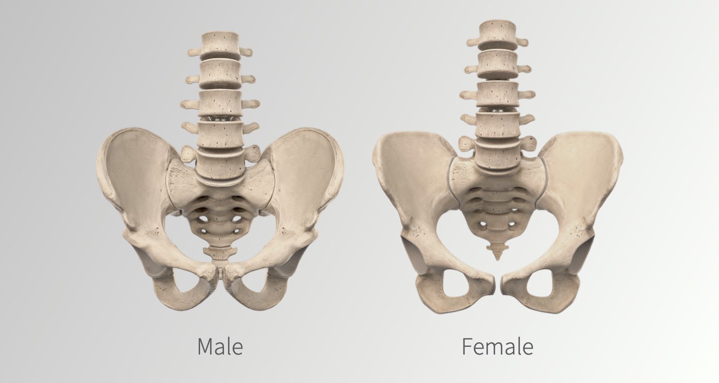
Choose whether to study principally on the female or male skeleton, with equal representation of both. The unique details that represent the female body such as the sexual dimorphic differences found in the pelvis, or finer details of the skull, have been carefully depicted.
From the subtle differences in the long bones to the extremely detailed skeletal parts, surfaces and landmarks of the pelvis, our uniquely female skeletal system is ready for stand-alone study or comparison against the male skeleton.
Fibers, tendons and musculature. All fully Female

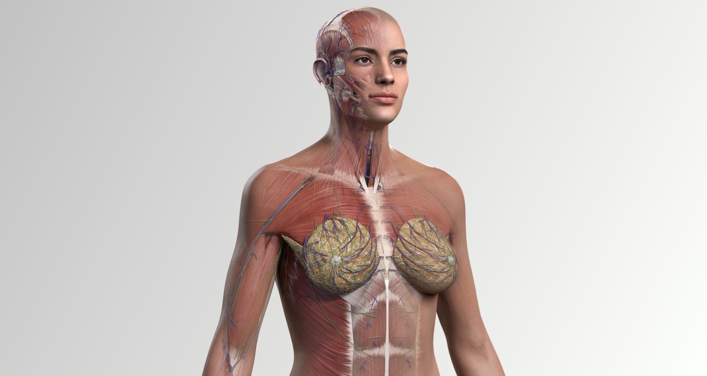
The new female model accurately reflects the correct muscle composition while retaining the clear and concise shape of each muscle to facilitate identification and understanding of form and function.
In order to do this while also creating an accurate representation of the female body, each muscle has been built to carefully represent what is commonly found amongst the female demographic. This has meant we have reduced the overall volume of muscle mass by about 30%, following the research from subject matter experts and academic studies.[3, 4]
A new perspective on female-specific regions

As proud as we are to represent the female body in full, standing shoulder-to-shoulder with our male counterpart, we have worked extra-hard on the defining female-specific regions of the body.
The breast tissue of the female model can be hemisected or quartered in-app to reveal the underlying tissues. Details such as the inferior distribution of the mammary glands, which we chose to represent as non-lactating, are representative of the majority of the population unlike most anatomical depictions to date.
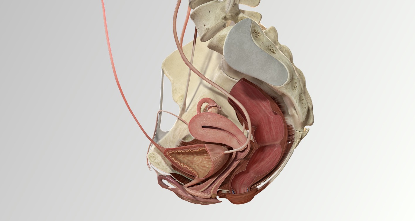
The reproductive organs from the internal and external genitalia have been remodeled to accurately show their continued relationship, such as the important contribution of the anterior vaginal wall to the vestibule. We have based the shape of the clitoris on the latest academic research so that it now presents in the classic J-crook shape and ensured to provide as much detail in the surrounding neurovasculature so that students can fully understand this complex region of the body.
It is through a better depiction of these often-misrepresented regions that we hope to provide better patient outcomes through better student education.
Male and female anatomy. Every difference. Every detail.

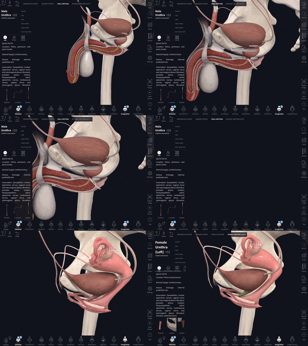
With one long-press of the Models Button, you can easily switch your model setup between the female and male models, making comparative anatomy teaching seamless.
Existing reference materials such as illustrations and physical models typically focus only on female-specific differences of the thoracic and pelvic areas. Using our comparative technology, you as an educator can easily switch the sex of any region in the body. This allows difficult comparisons, such as those of the pelvic area, to be better communicated through presenting them from multiple angles or in closer detail.
We are incredibly excited to take this step forward to improved representation within the field of anatomy and this is only the first of many projects we will be working on over the next few years that address the under-representation of diverse groups within anatomy. We hope you enjoy using our new full female model, which we are certain will create a lasting difference for educational equality.
A full-female model exclusive, that makes mastering anatomy more inclusive.
References
- https://papers.ssrn.com/sol3/ papers.cfm?abstract_id=383803
- https://www.healthline.com/ health-news/ we-dont-have-enough-women-in-clinical-trials-why-thats-a-problem
- Gallagher D, Heymsfield SB. Muscle distribution: variations with body weight, gender, and age. Applied Radiation and Isotopes. 1998;49(5-6):733-734.
- Janssen I, Heymsfield SB, Wang, Z, Ross R. Skeletal muscle mass and distribution in 468 men and women aged 18-88 yr. Journal of Applied Physiology. 2000; 89:81- 88.
