The small intestine is a key part of the digestive system. Spanning 6-7m within the abdominal cavity, the small intestine has roles in digestion and absorption.
The small intestine is divided into three parts: the duodenum, the jejunum and the ileum. The first part, the duodenum, is the shortest section. It is about 25-30cm long. It starts at the pylorus of the stomach and forms a C-shape around the head of the pancreas. The duodenum is a site of digestion as well as absorption. For instance, bile is released into the duodenum to emulsify fats. Pancreatic enzymes are also released to digest proteins, carbohydrates and fats.
The second part of the small intestine is the jejunum. The jejunum continues from the duodenum up to the ileum. It is about 2.5-3m long and is coiled within the abdominal cavity. Absorption mainly takes place here.
The final part of the small intestine is the ileum. The ileum continues from the jejunum and ends at its connection to the large intestine. It coils within the abdominal cavity for about 3.5-4m. The ileum is also a site of absorption. Absorption is a key process in digestion as it takes the nutrients and water from food so they can be used in the body for other processes.
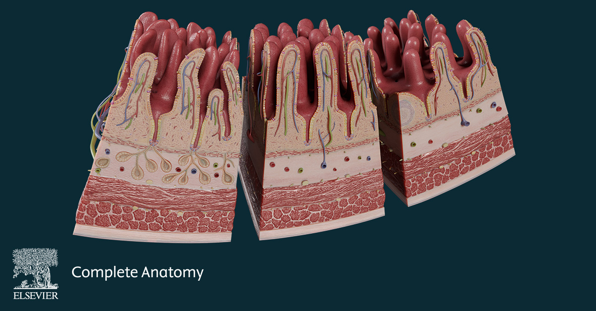
The mucosa of the small intestine is lined with threadlike projections called villi. The villi increase the surface area for absorption of fluids and nutrients. In the duodenum, the villi are leaf-shaped and gradually become more finger-like in the ileum. Within each villus is a central lymph vessel called a lacteal. Lacteals are important for the absorption of lipids. A submucosal vascular plexus is also found within the villi. This is a network of intercommunicating blood vessels which absorb products of digestion such as vitamins, minerals and water.
The lining of the small intestine is made up of enterocytes. These are simple, tall columnar epithelial cells. The primary function of enterocytes is absorption. The apical surface is covered in microvilli which further increase the surface are for absorption.
Goblet cells are mucous glands found in the epithelium of the small intestine. These glands secrete mucus which protects and lubricates the surface of the intestines. Enteroendocrine cells are also found in the epithelium. Their secretions have a number of functions including affecting gastrointestinal motility, pancreatic and biliary secretions, and gastrointestinal epithelial growth.
Check out the new Small Intestine Detailed Model in Complete Anatomy to learn more!