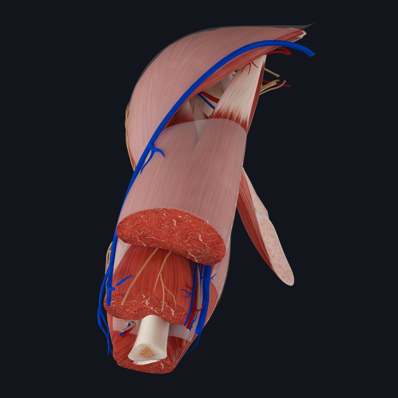
The muscles of the limbs are divided into discrete compartments. Over the course of the next few weeks we will learn about each of these individual collections of muscle in detail and delve into the benefits and drawbacks of having such segregation.
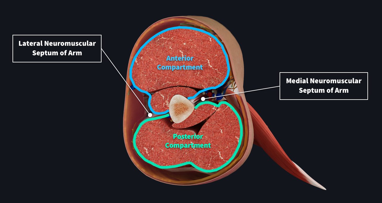
The upper limb is divided into three regions: the hand, the arm, and the forearm. The two proximal regions are further anatomically divided into two compartments, namely anterior and posterior. In addition to the bony framework, an impressive organization of deep fascia forms the divisions of these compartments.
In the arm, emerging from the centrally located humerus, are two folds of tough connective tissue, one travelling medially towards the midline and the other progressing laterally until it reaches the superficial fascia. These two intermuscular septa form the shared boundary between the anterior and posterior compartments of the arm. As they reach the superficial tissue, this dense connective tissue divides to form a sleeve around the collection of muscles located anterior and posterior to the bone. Often this fascia will also encapsulate neurovasculature in addition to the large muscle bellies.

In the arm, the anterior compartment contains long and short heads of biceps brachii, brachialis and coracobrachialis muscles, all of which are innervated by the musculocutaneous nerve and supplied by the brachial artery. The posterior compartment is home to long, short and medial heads of triceps brachii muscle as well as anconeus. The radial nerve travels within the posterior compartment proximally before it pierces the lateral intermuscular septum at the origin of brachioradialis. The rest of the neurovasculature in the arm is embedded in superficial fascia on the medial side of the humerus, outside the two named compartments.
Get a full understanding of the different muscle compartments with the world’s most advanced 3D anatomy platform, complete with learning views such as Muscle Innervation and Arterial Supply.
