This month, we’re launching an exciting new addition to the platform: Cell Biology. Transform how you understand the minute cellular workings of the body with the first in a brand-new upcoming series of detailed anatomy models. Experience unparalleled levels of detail in the Blood, Sperm and Secondary Oocyte models.
Blood
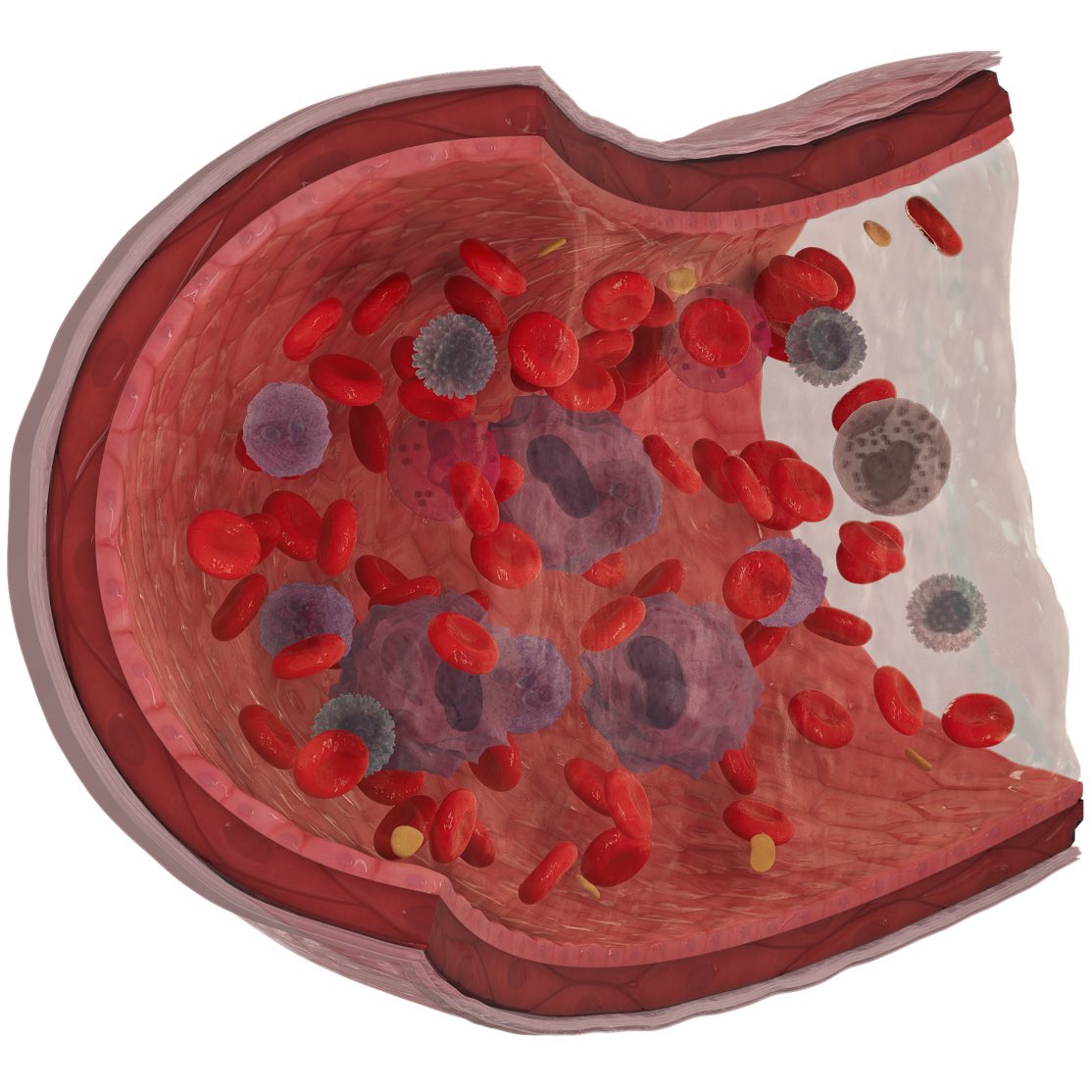
The Blood model offers an insight into a blood vessel and the composition of blood found within it.
Journey through the various tunica layers of an arteriole and into the plasma containing erythrocytes, leukocytes and platelets.
Students can compare the different types of cells that make up blood. See incredibly small platelets rolling along the edges of the vessel, compared to the largest blood cell, monocyte, found suspended in the plasma.
Each of the uniquely different leukocytes are represented, and can be distinguished by their individual nuclei, as well as their shape and surface morphology.
Sperm
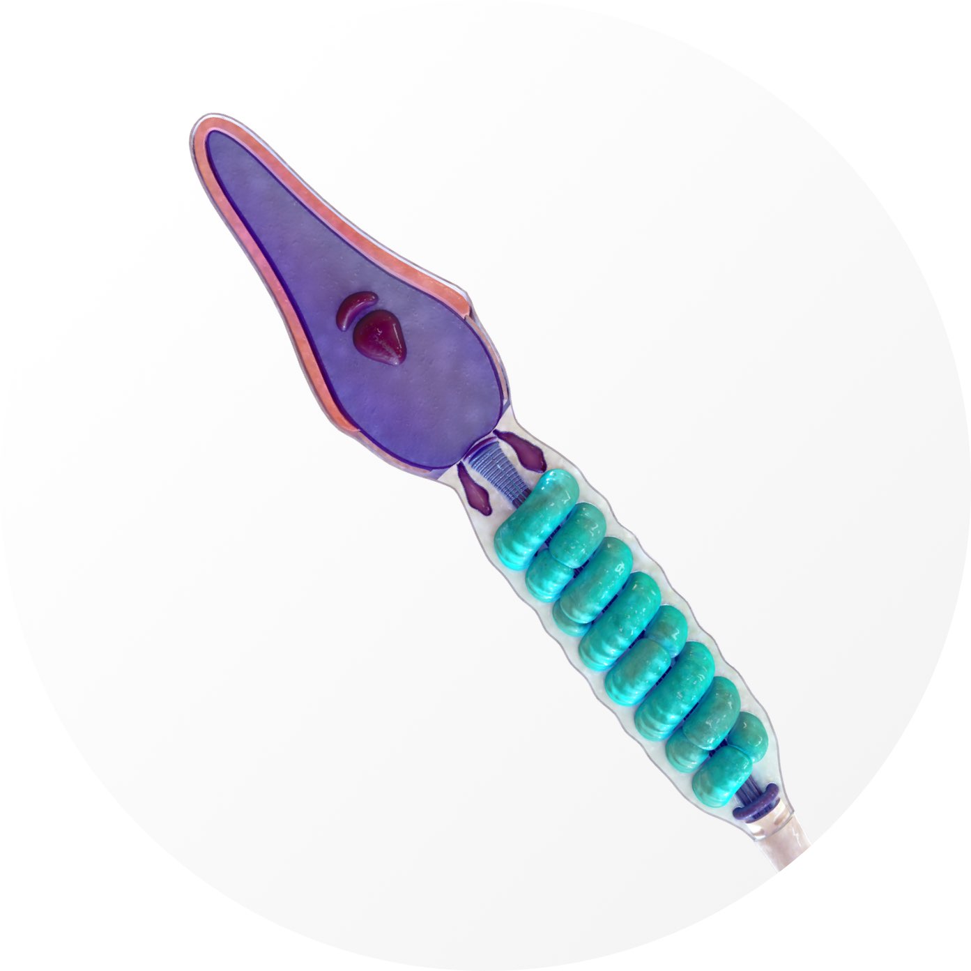
This microscopic Sperm model allows students to visualize the smallest cell in the human body, the sperm cell. The tiny size of this cell makes it incredibly difficult to study. However, with this 3D model, the variety of structures and their functions can be truly appreciated.
Navigate through a sperm cell by layer and by region. Explore the head region by uncovering the associated plasma membrane, cytoplasm, acrosome, nuclear envelope and so on.
Every minute detail has been considered, from the centrosome within the striated column in the neck, to the fibrous sheath running the length of the tail.
Secondary oocyte
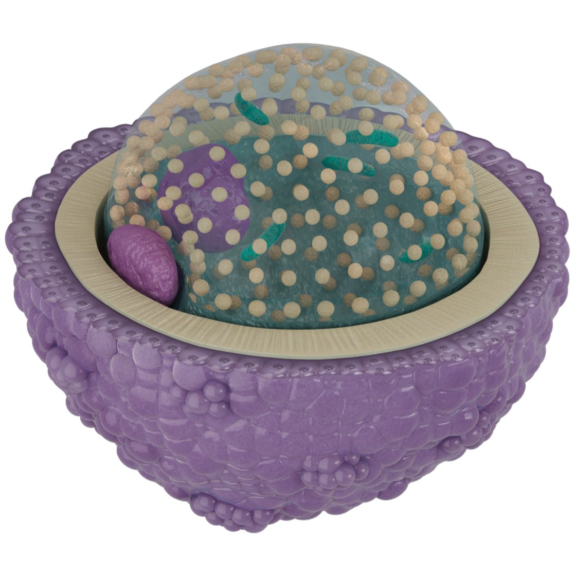
The secondary oocyte is an immature egg cell. It is released during ovulation and is the form of the egg cell which fuses with sperm cells. It is therefore an exceptional comparison model when studied alongside the sperm.
Investigate the layers that make up the secondary oocyte, such as the corona radiata and zona pellucida, and how they contribute to successful fertilization processes.
Navigate your way down to the very centre of the oocyte, where the cortical granules, mitochondria and nucleus can be seen suspended in the jelly-like cytoplasm.
We’re releasing an update to include these 3 new detailed models very soon, and they will be accessible as part of a Student Plus, Educator or Professional license. If you haven’t already done so, sign up today to Try Complete Anatomy for FREE for 3 days and experience the power of 3D anatomy learning.