Complete Anatomy 2020 features 15 microanatomy models that explore the most minute details of the human anatomy, right down to the cellular level.
The comprehensive and ever-expanding collection of microanatomy models is designed to take you on a journey from the gross anatomy, right down to the cellular level, with everything in between. Explore a skeletal striated muscle in the gross model, then see its fibres in even more detail in a dedicated microanatomy model.
Peripheral nerve
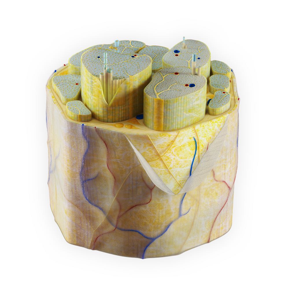
We’ve designed a peripheral nerve model which clearly shows the organization of nerve tissue within primary and secondary fasciels, surrounded by perineurium and epineurium. On a deeper level we have represented four different kinds of nerve fibers, general visceral efferent nerve fibers, somatic motor efferent nerve fibers, general visceral afferent nerve fibers and finally somatic sensory afferent nerve fibers and the differences between each such as size, position, myelin sheath and more.
Skeletal striated muscle
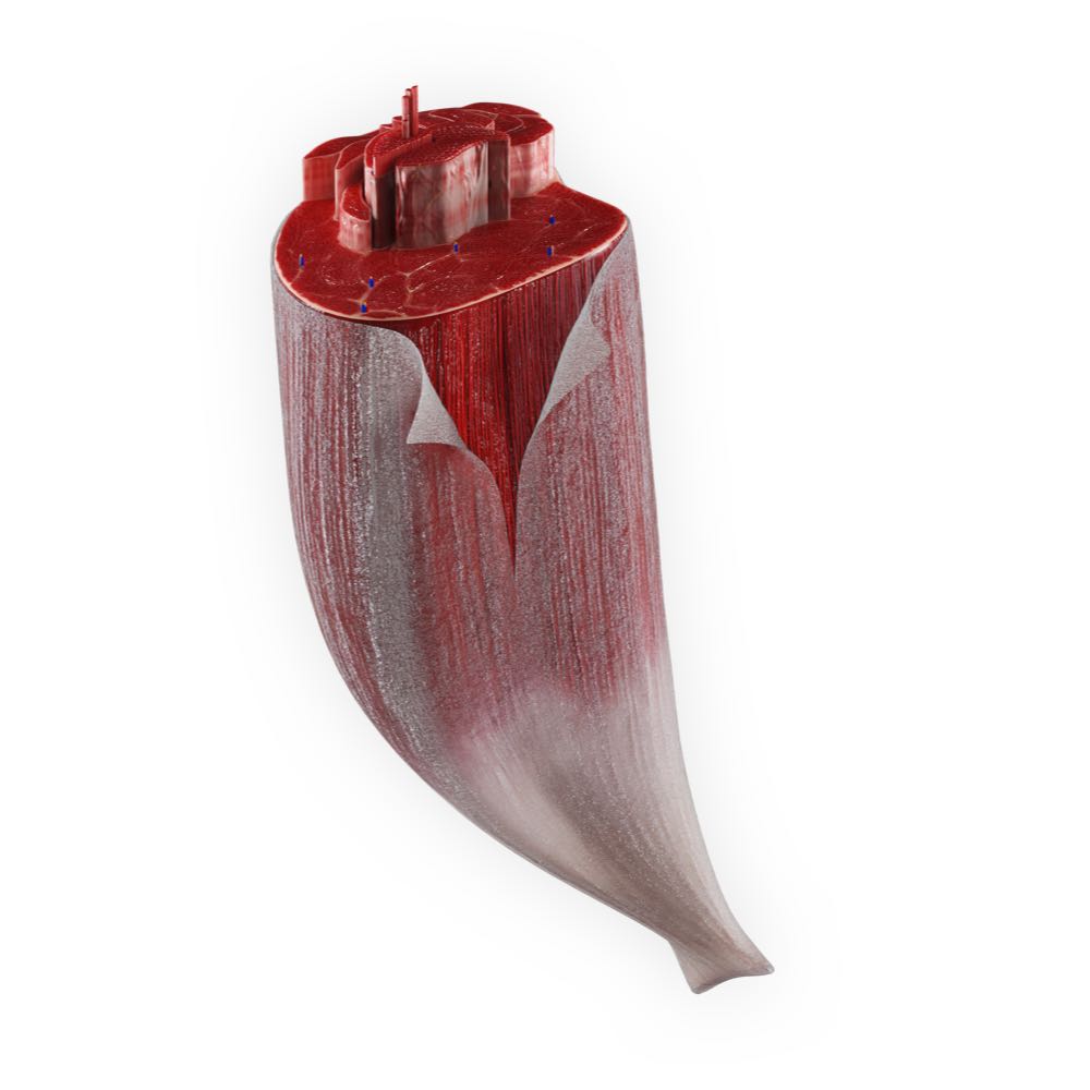
This model takes the belly of the long head of biceps femoris and section it to show the organization of muscle fasicles, right down to the extrafusal muscle fiber, and the surrounding organisation of connective tissue and vasculature.
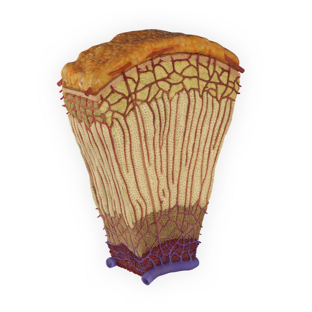
Bone Tissue
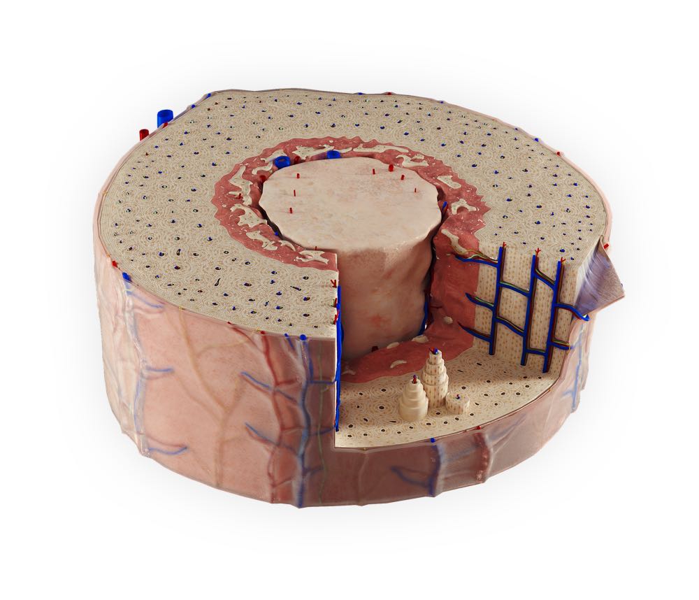
The bone tissue model represents a cross-section of a long bone. Explore the layers of the periosteum, the arrangement of osteons and how the blood supply penetrates through the bone to reach the medullary cavity filled with yellow bone marrow.
Skin
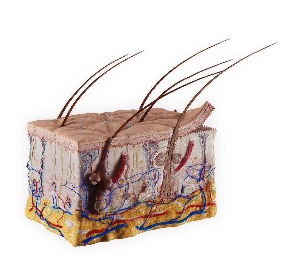
Reminiscent of the classical section view found in many textbooks, our Skin microanatomy model is designed using histological and scanning electron microscope data adding a layer of accuracy that you won’t find anywhere else! Of the many structures found within a section of the skin, users can explore the hair follicle in its entirety or in cross-section where they can study the makeup of hair starting from it’s germinal matrix, the relationship of sebaceous glands, the arrector pili muscle and surrounding cellular tissue which together form the pilosebaceous unit.
Blood vessels
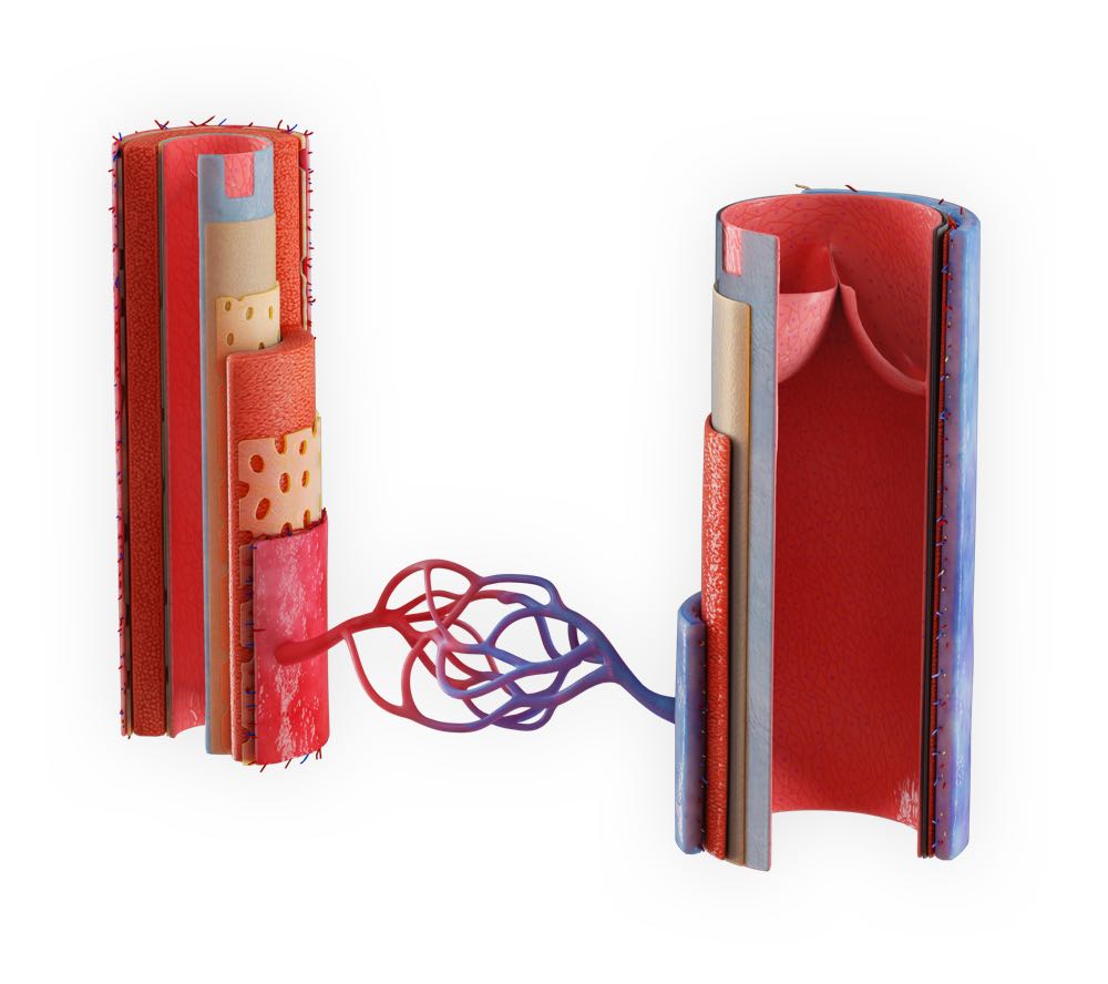
A comparative model showing the key structural differences between an artery and vein such as the differences in the size and quantity of their layers. For example, explore the external and internal elastic membrane found within an artery and how it also contains smooth muscle.
Lymph Node
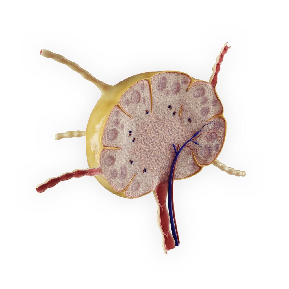
The lymph node is composed of different layers, the cortex, paracortex and the medulla. Explore the different types of cells found in each layer such as the B-lymphocytes which form lymphoid nodules. The model also represents the afferent and efferent flow of lymph and the difference present in each type of vessel.
Bronchial tree
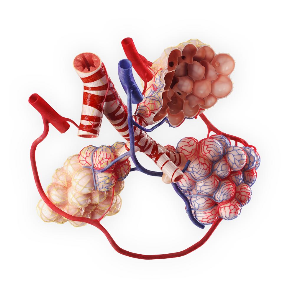
This model takes the bronchial tree and the alveoli, key functional units of the lungs and shows them in high detail, such as the relationship of the spiral network of smooth muscle cells, the elastic fibers around the alveolar ducts and saccules. The model also represents the dual blood supply of oxygenated and deoxygenated blood.
Skeletal muscle fiber
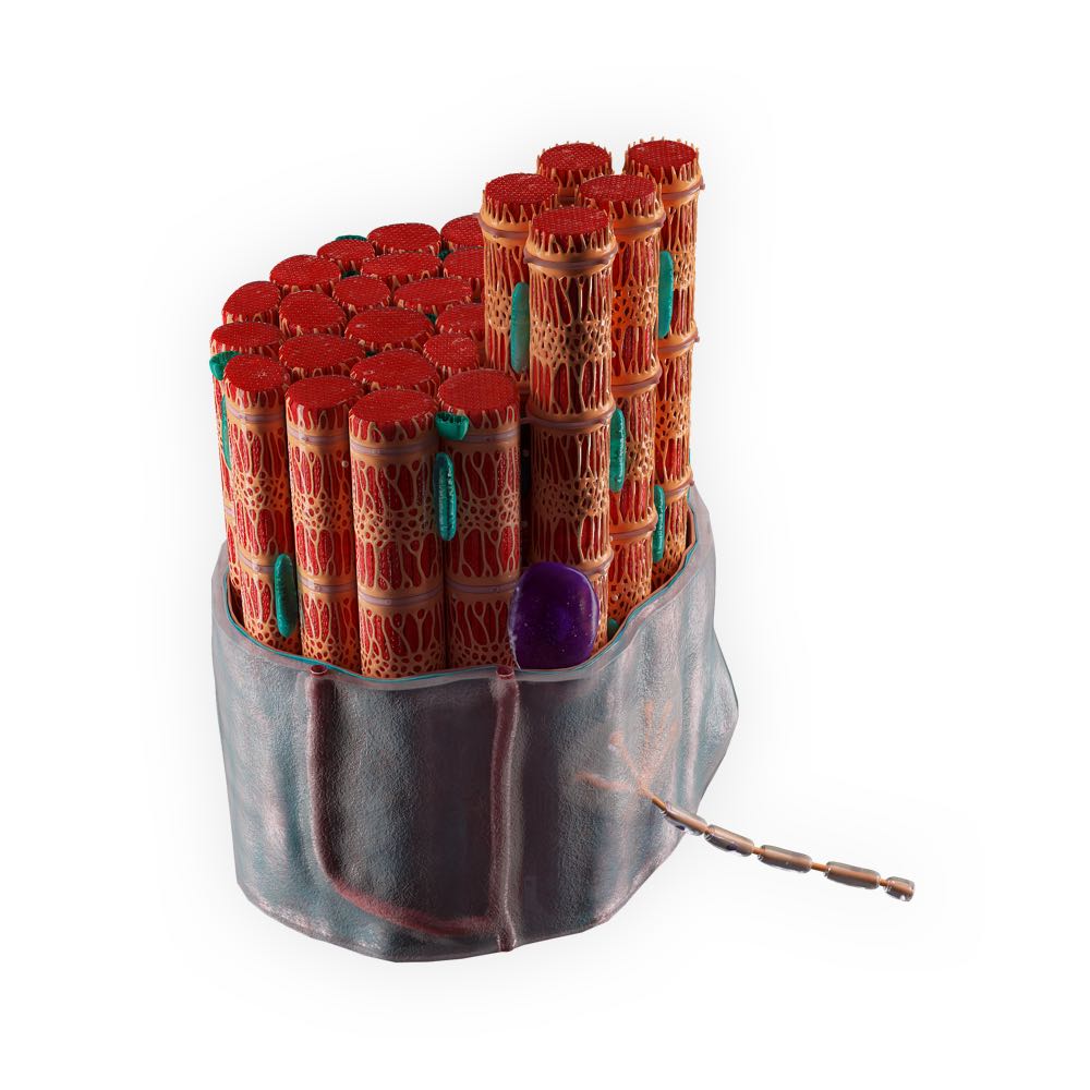
Following on from the skeletal striated muscle model we then go deeper, showing a detailed breakdown of a muscle fiber and how it is composed of myofibrils, their composition of actin and myosin and the supporting structures around them such as mitochondria, T tubules, and the layers of endomysium and sarcolemma. The model also represents a neuromuscular junction and its relationship with the myofibrils.
Tooth
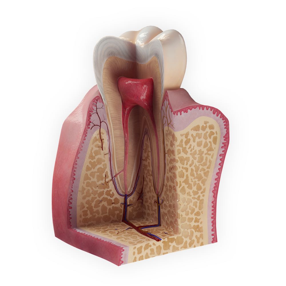
Ideal for any student studying dentistry, this model displays a sectioned view of a molar tooth, revealing the inner layers of the enamel and dentine along with texture details such as the enamel spindles, Striae of Retzius and enamel prisms. Go deeper into the pulp and view the blood and nervous supply to the tooth and how these structures pass through the root and alveolar bone. Note the important relationship of the different layers that make up surrounding gingiva such as the epithelium, lamina propria and dentogingival fibers.
Tendon

Similar to the striated muscle model, the tendon model shows users the organization of this tissue into tertiary, secondary and primary fiber bundles of collagen. Again we represent the correct blood supply as well as connective packing such as the endotendon and epitendon.
Kidney Lobe
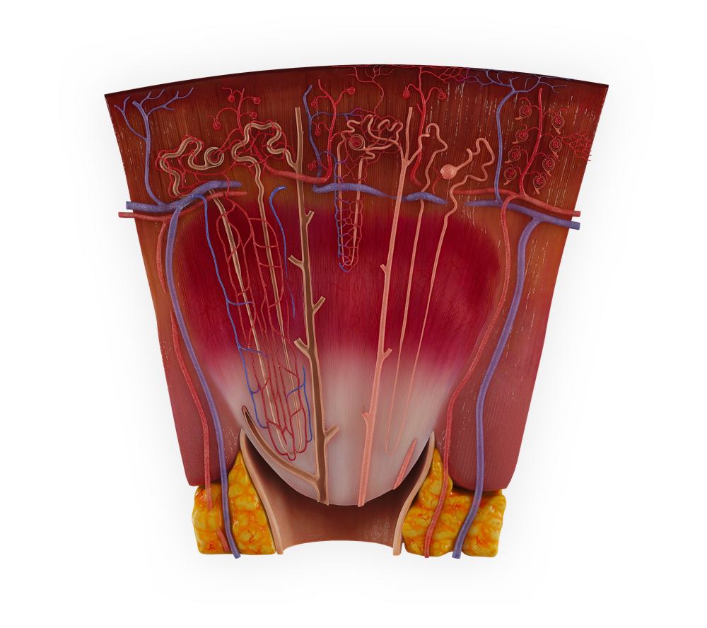
Examine the different kinds of the nephron, the juxtamedullary nephron and the cortical nephron. Students will also be able to explore the classic breakdown of the nephron into the proximal convoluted tubule, the descending and ascending limb of the loop of Henle, the distal convoluted tubule and eventually how they lead to the collecting duct which empties into the renal papilla.
The Eye
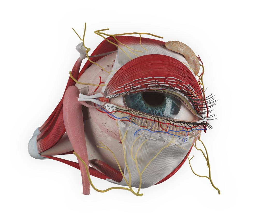
The detailed prosection of the eye shows a mixture of gross anatomy and detailed microanatomy. Explore the relationship of the eye to other structures found within the orbit such as the extraocular muscles and lacrimal apparatus. Remove the layers of the eyelid to reveal the neurovasculature, the tarsal plate, tarsal sebaceous glands and more. Create a sagittal cross section to reveal the lens and its relationship to the ciliary ring and ciliary muscles. The retinal layer is also exposed and shows a detailed view of the fovea and macula.
Tongue Section
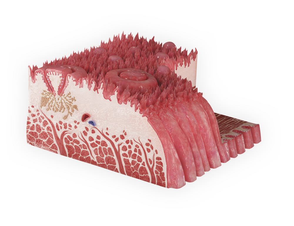
Get a detailed look at the different kinds of papillae, filiform, vallate, fungiform and foliate. The model also represents their unique taste buds and relationship with the sensory nerve fibers which carry the action potential needed to register taste.
Suprarenal gland
View the cellular break down of the suprarenal gland and how it is organised into layers and explore each layers function. Pay particular attention to the detailed blood supply which is vitally important for transporting the hormones that the suprarenal gland produces.
Olfactory Organ
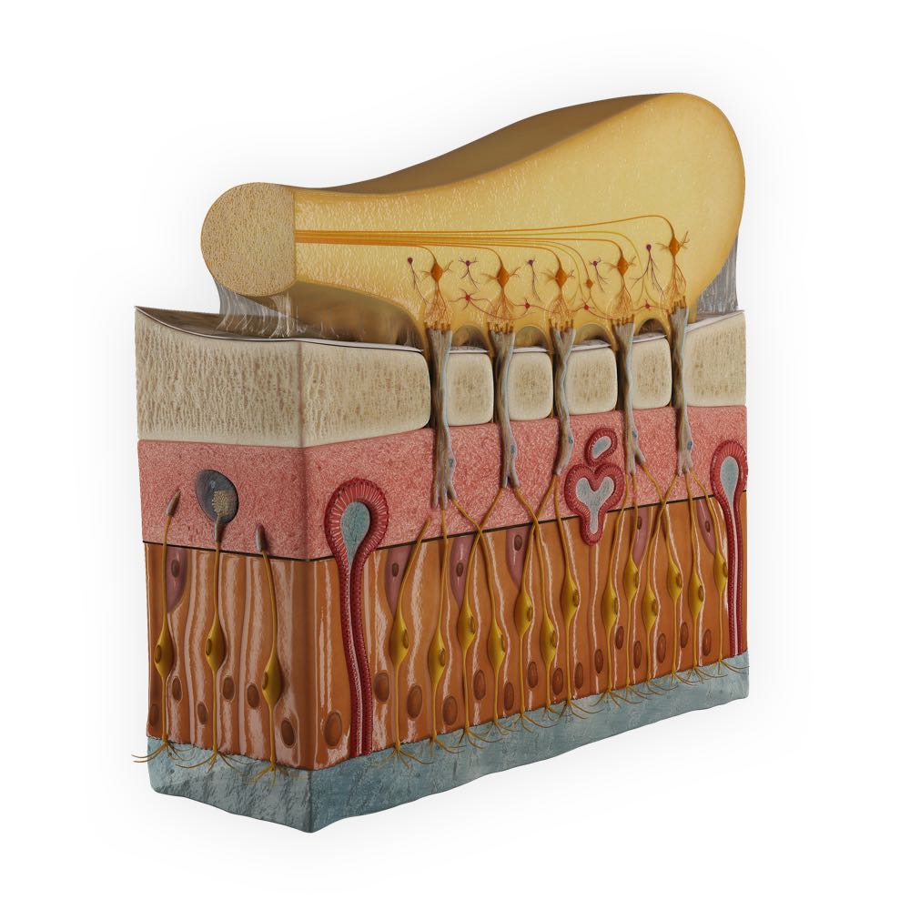
A detailed view of the olfactory mucosa and the olfactory bulb. View the organization of the olfactory epithelium, how it is made up of a combination of the olfactory neurons, supporting cells and basal cells. Follow how an odour molecule generates an action potential required for the sense of smell, from the olfactory neuron and it’s cilia spreading out onto the surface of the olfactory epithelium. The dendrite leading to the cell body situated halfway in the olfactory mucosa, between the supporting cells. The axon and how they group together in ensheathing cells to eventually reach the olfactory bulb. Even the neural pathways within the olfactory bulb are represented, for students to study in a clear and concise manner.
Unlock a whole new world of anatomy & physiology today. Get full access to ALL 14 microanatomy with a FREE 3-day trial of Complete Anatomy. Try it for free today.