For the final instalment of our examination of the muscular compartments let’s dive into the divisions of the leg. As we said before, the leg is divided into three muscular compartments, however this can sometimes be classified as four muscle groups. The anterior and lateral compartments occupy only a small portion of the muscular volume of the leg. The beefy posterior compartment is a major contributor to our mobility, specifically in walking and running, and is thus composed of large muscles – these muscles can be divided into deep and superficial groups.
The anterior compartment, also known as the extensor compartment, contains muscles that dorsiflex or extend the foot at the ankle joint, and muscles that flex the toes. They are tibialis anterior, extensor digitorum longus, extensor hallucis longus, and fibularis tertius. Also within this compartment is the anterior tibial artery and vein and the deep fibular nerve.
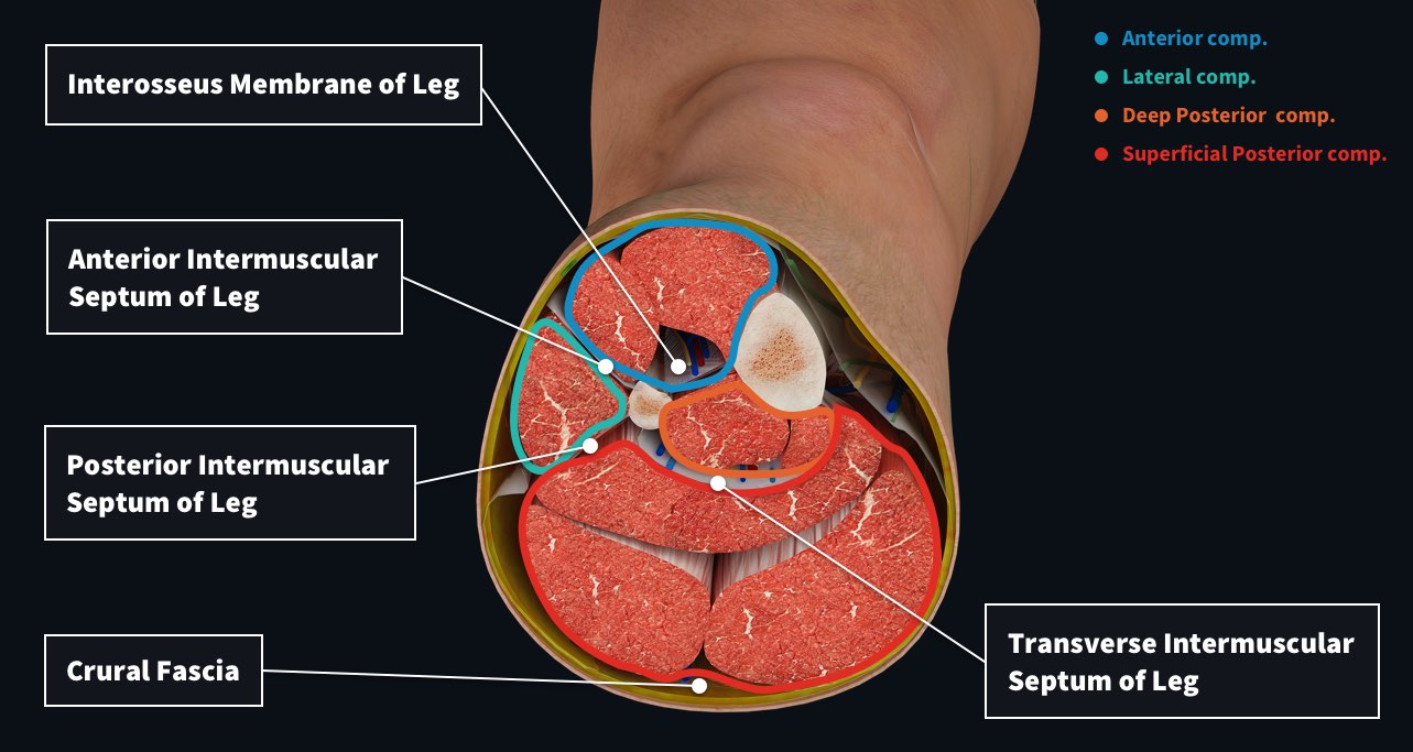
Within the lateral compartment are muscles that are primarily involved with eversion of the foot, to tilting the sole of the foot laterally. These two muscles are fibularis longus and brevis, and their innervating nerve, the superficial fibular nerve. Dividing this muscle group from the anterior compartment is the anterior intermuscular septum, and separating it from the posterior compartment is the posterior intermuscular septum. Pretty straightforward, right?
The muscles of the large posterior group are primarily responsible for plantar flexion of the foot, a movement that allows us to stand on our tippy toes and an essential part of gait. These muscles essentially lift the entire body weight on contraction. The superficial group comprises three muscles which are the major contributors to plantar flexion – they are soleus, gastrocnemius, and plantaris. Muscles in the deep posterior group are involved with flexion of the toes as well as stabilization of the knee and arch of the foot, and are called flexor digitorum longus, flexor hallucis longus, tibialis posterior and popliteus. Alongside these muscles are the posterior tibial artery and vein, the fibular artery and vein and the tibial nerve, which provides innervation to all of the muscles in the posterior compartment.
The transverse intermuscular septum indicates the division between deep and superficial muscles groups and the deep group is separated from the anterior compartment by the tibia, fibula, and the interosseous membrane running between them. Surrounding the entire leg is deep crural fascia. As it travels around the leg it envelops the external surface of the muscle compartments and blends with the periosteum overlying the tibia.
Use the power of 3D anatomy to better understand the muscle compartments of the anatomy. To see the learning power of Complete Anatomy, try it for FREE today.