Let’s get into the guts of our new detailed model. The large intestine derives its name from the fact that its diameter is noticeably larger than that of the small intestine. It is made up of the cecum, colon and rectum.
The colon is further divided into the ascending, transverse, descending and sigmoid colons. The latest detailed model in CA takes a closer look at the Transverse Colon allowing users to explore this section of the large intestine in more detail. The transverse colon travels horizontally across the abdomen below the liver, stomach and spleen. This part of the colon sits above the small intestine and extends from the hepatic flexure to the splenic flexure, the points at which the colon bends at a 90° angle.
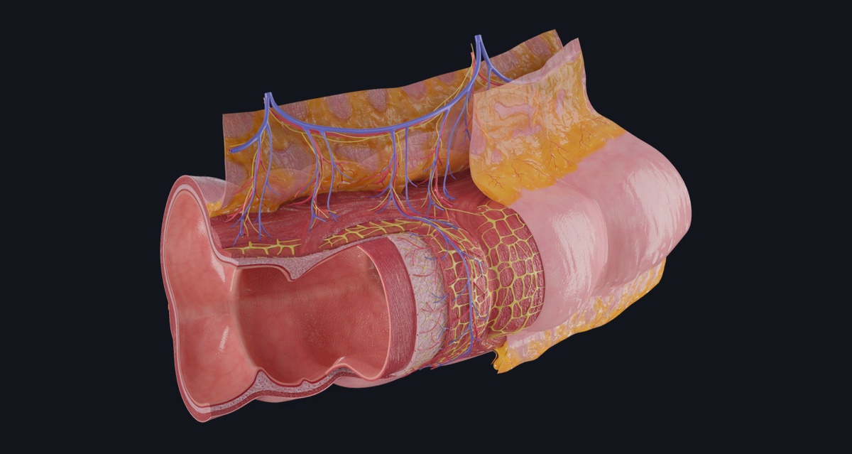
The Transverse Colon model will allow users to explore the following layers of the colon in even more detail than is possible in the gross model:
- Mucosa— The mucosa is the inside layer of the gastrointestinal tract which faces the lumen. The mucosa itself is composed of three separate layers—the inner mucous-lined epithelium, the lamina propria made up of loose fibrous connective tissue, and the muscularis mucosae—a thin smooth muscle layer.
- Submucosa— The submucosa of the alimentary canal is made up of layers of irregular dense connective tissue. Many small glands, blood vessels and parasympathetic nerves of the submucosal plexus are found within these layers.
- Muscular Layer— The muscular layer is a thick layer of smooth muscle tissue that lies between the mucosa and serosa and wraps around the submucosa. The muscular layer is composed of an inner circular layer ⭕ and outer longitudinal layer 🔛. In the colon, longitudinal muscle aggregate into tape-like strips known as tenia coli. Like the submucosa, the muscular layer contains nerves organized into a plexus called the myenteric plexus which lies between the two muscle layers.
- Serosa— The serosa is the outermost layer of the gastrointestinal wall where it has a protective function. It consists of mesothelium lying on a subserosal connective tissue layer.
You can explore all of this and more in Complete Anatomy, try it for FREE today.