Today let’s learn about the dura mater while also getting an exclusive look of our revamped Head & Neck model, coming this summer to Complete Anatomy.
Dura mater is the outermost layer of the meninges that cover our brain. It lies underneath the bones of the skull and vertebral column. It is a thick and very tough material that protects the brain. ?.
Dura mater is the outermost layer of the meninges that cover our brain. It lies underneath the bones of the skull and vertebral column. It is a thick and very tough material that protects the brain. ?.
The dura within our cranial cavity contains two sheets of connective tissue:
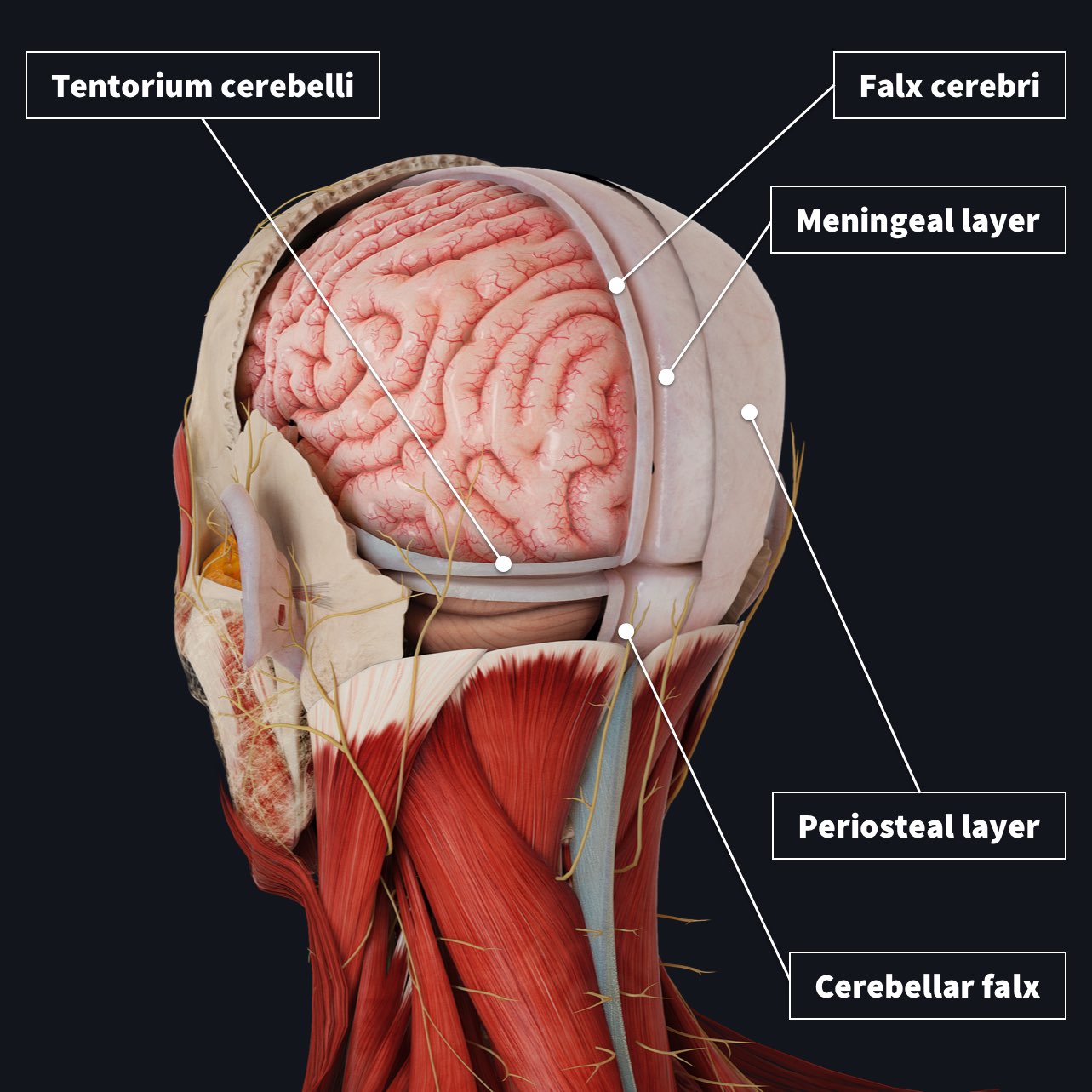
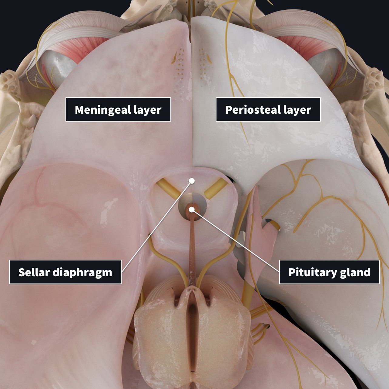
- Periosteal layer- covers inner surface of the skull
- Meningeal layer- deep to periosteal layer within cranial cavity. The only layer present in vertebral column.
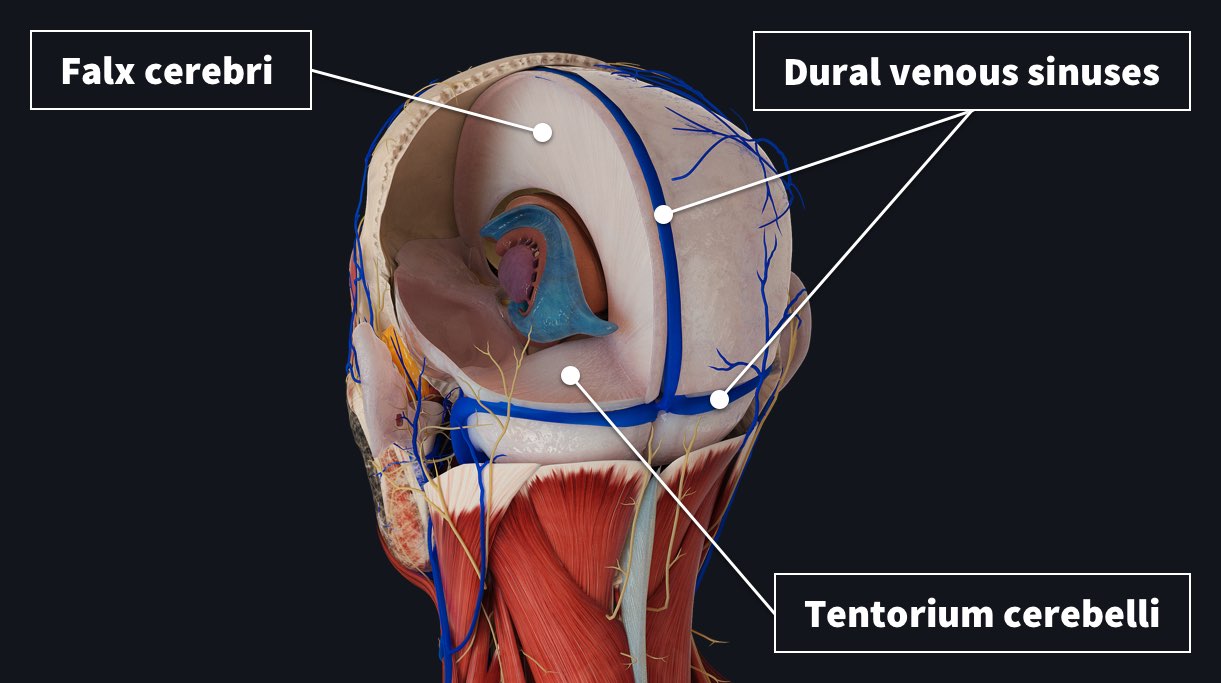
The dural venous sinuses are located between these two layers. They allow the venous vasculature of the cranium to drain into the internal jugular veins ?.
Within some areas of the skull, the meningeal layer of dura mater folds inwards as a dural reflection. These folds help to partition the brain and divide the cranial cavity into several compartments.
For example…
↕️ Tentorium cerebelli- divides the cranial cavity into supratentorial and infratentorial compartments. It covers the top of the cerebellum and supports the occipital lobes.
↔️ Falx cerebri- a large crescent shaped fold projecting down into the longitudinal fissure, separating right and left cerebral hemispheres.
Two other dural reflections are..
↔️ Cerebellar falx- a vertical fold that lies inferior to the tentorium cerebelli and the posterior part of the posterior cranial fossa. It partially separates the cerebellar hemispheres.
⏺ Sellar diaphragm- the smallest dural fold, a circular sheet of dura suspended between clinoid processes, forming a partial roof over the hypophysial fossa. Covers the pituitary gland in the fossa and allows passage for pituitary stalk.
There are many contributions to the blood supply and innervation of the dura mater, however it mainly receives its own vasculature from the middle meningeal artery and vein. It is innervated by the trigeminal nerve (V1, V2 and V3), which you can learn more about in this Anatomy Dissected video!